Loading
PDBj
MenuPDBj@FacebookPDBj@TwitterPDBj@YouTubewwPDB FoundationwwPDB
RCSB PDBPDBeBMRBAdv. SearchSearch help

신종 코로나바이러스 특집 콘텐츠

이 페이지는 다른 언어로도 제공됩니다: English日本語简体中文繁體中文
코로나 바이러스
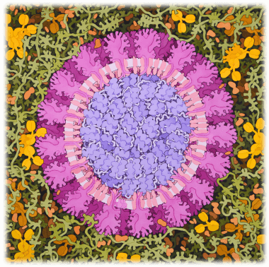
코로나 바이러스, 2020. David S. Goodsell @ RCSB PDB에 의한 오리지널 일러스트를 수정

신종 코로나바이러스 감염증 (Novel Coronavirus disease 2019:COVID-19)의 확산이 세계적으로 매우 큰 문제가 되고 있습니다. 시급히 새로운 바이러스(Severe Acute Respiratory Syndrome Coronavirus 2: SARS-CoV-2)를 이해하고 효과적인 약을 개발하기 위해, 신속하게 구조 생물학 연구가 시작되고 있습니다. PDBj는 사용자의 편의를 도모하기 위해 신종 코로나바이러스의 특집 페이지를 제공하고 있습니다. 매주 수요일에 최신 항목이 추가됩니다.

바이러스 단백질에 관한 해설기사는 아래의 「이달의 분자」 페이지를 참조하십시오.

「전체 엔트리」 탭에는, 모든 PDB ID가 포함되어 있습니다. 이용자가 전체의 독립된 엔트리를 검색하고 싶을 때 사용하실 수 있으며, 동일 저자에 의한 그룹등록의 엔트리도 전부 구별되어 있습니다. 「대표구조」 탭에는, 분해능이 가장 높은 대표구조만을 표지하고 있습니다. 아미노산 서열이 100 % 일치하는 PDB 엔트리의 경우, 다른 리간드를 포함하여도 대표구조에는 제외됩니다. 「최신 엔트리」 탭은, 이번주 공개된 가장 최근의 엔트리 입니다.


Created: 2020-09-03 (last edited: more than 1 year ago)2022-09-02
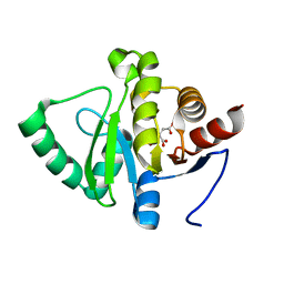
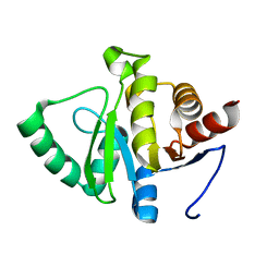
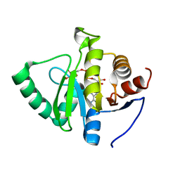
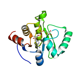
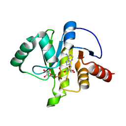
7TWH
DownloadVisualize
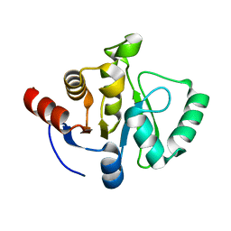
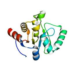
BU of 7twh by Molmil
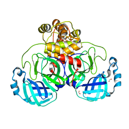
Crystal structure of SARS-CoV-2 NSP3 macrodomain at 293 K (P43 crystal form, 290 kGy)
Descriptor: Non-structural protein 3
Authors:Correy, G.J, Fraser, J.S.
Deposit date:2022-02-07
Release date:2022-02-23
Last modified:2023-10-18
Method:X-RAY DIFFRACTION (1.1 Å)
Cite:The mechanisms of catalysis and ligand binding for the SARS-CoV-2 NSP3 macrodomain from neutron and x-ray diffraction at room temperature.
Sci Adv, 8, 2022
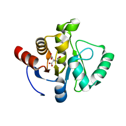
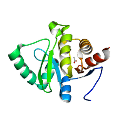
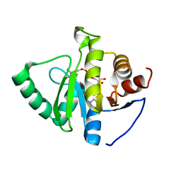
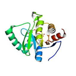
7TWI
DownloadVisualize
BU of 7twi by Molmil
Crystal structure of SARS-CoV-2 NSP3 macrodomain at 293 K (P43 crystal form, 539 kGy)
Descriptor: Non-structural protein 3
Authors:Correy, G.J, Fraser, J.S.
Deposit date:2022-02-07
Release date:2022-02-23
Last modified:2023-10-18
Method:X-RAY DIFFRACTION (1.1 Å)
Cite:The mechanisms of catalysis and ligand binding for the SARS-CoV-2 NSP3 macrodomain from neutron and x-ray diffraction at room temperature.
Sci Adv, 8, 2022
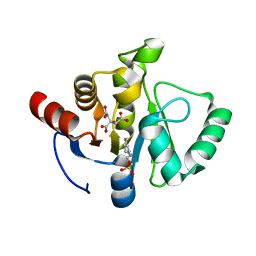
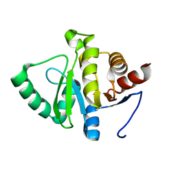
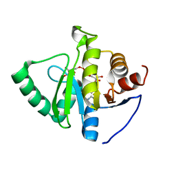
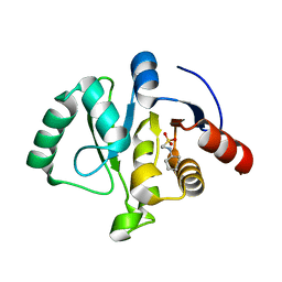
7TWJ
DownloadVisualize
BU of 7twj by Molmil
Crystal structure of SARS-CoV-2 NSP3 macrodomain at pH 4 (P43 crystal form)
Descriptor: CITRIC ACID, Non-structural protein 3
Authors:Correy, G.J, Fraser, J.S.
Deposit date:2022-02-07
Release date:2022-02-23
Last modified:2023-10-18
Method:X-RAY DIFFRACTION (0.9 Å)
Cite:The mechanisms of catalysis and ligand binding for the SARS-CoV-2 NSP3 macrodomain from neutron and x-ray diffraction at room temperature.
Sci Adv, 8, 2022
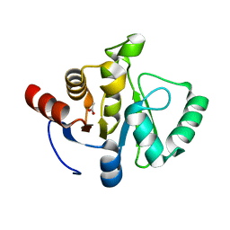
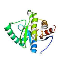
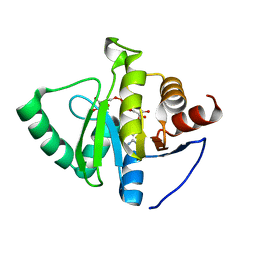
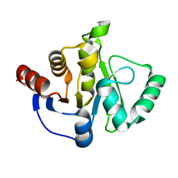
7TWN
DownloadVisualize
BU of 7twn by Molmil
Crystal structure of SARS-CoV-2 NSP3 macrodomain at pH 5 (P43 crystal form)
Descriptor: 2-[N-CYCLOHEXYLAMINO]ETHANE SULFONIC ACID, CITRIC ACID, Non-structural protein 3
Authors:Correy, G.J, Fraser, J.S.
Deposit date:2022-02-07
Release date:2022-02-23
Last modified:2023-10-18
Method:X-RAY DIFFRACTION (0.9 Å)
Cite:The mechanisms of catalysis and ligand binding for the SARS-CoV-2 NSP3 macrodomain from neutron and x-ray diffraction at room temperature.
Sci Adv, 8, 2022
7TWO
DownloadVisualize
BU of 7two by Molmil
Crystal structure of SARS-CoV-2 NSP3 macrodomain at pH 6 (P43 crystal form)
Descriptor: 2-[N-CYCLOHEXYLAMINO]ETHANE SULFONIC ACID, CITRIC ACID, Non-structural protein 3
Authors:Correy, G.J, Fraser, J.S.
Deposit date:2022-02-07
Release date:2022-02-23
Last modified:2023-10-18
Method:X-RAY DIFFRACTION (0.9 Å)
Cite:The mechanisms of catalysis and ligand binding for the SARS-CoV-2 NSP3 macrodomain from neutron and x-ray diffraction at room temperature.
Sci Adv, 8, 2022
7TWP
DownloadVisualize
BU of 7twp by Molmil
Crystal structure of SARS-CoV-2 NSP3 macrodomain at pH 7 (P43 crystal form)
Descriptor: ACETATE ION, Non-structural protein 3
Authors:Correy, G.J, Fraser, J.S.
Deposit date:2022-02-07
Release date:2022-02-23
Last modified:2023-10-18
Method:X-RAY DIFFRACTION (0.9 Å)
Cite:The mechanisms of catalysis and ligand binding for the SARS-CoV-2 NSP3 macrodomain from neutron and x-ray diffraction at room temperature.
Sci Adv, 8, 2022
7TWQ
DownloadVisualize
BU of 7twq by Molmil
Crystal structure of SARS-CoV-2 NSP3 macrodomain at pH 9 (P43 crystal form)
Descriptor: Non-structural protein 3
Authors:Correy, G.J, Fraser, J.S.
Deposit date:2022-02-07
Release date:2022-02-23
Last modified:2023-10-18
Method:X-RAY DIFFRACTION (0.9 Å)
Cite:The mechanisms of catalysis and ligand binding for the SARS-CoV-2 NSP3 macrodomain from neutron and x-ray diffraction at room temperature.
Sci Adv, 8, 2022
7TWR
DownloadVisualize
BU of 7twr by Molmil
Crystal structure of SARS-CoV-2 NSP3 macrodomain at pH 8 (P43 crystal form)
Descriptor: ACETATE ION, Non-structural protein 3
Authors:Correy, G.J, Fraser, J.S.
Deposit date:2022-02-07
Release date:2022-02-23
Last modified:2023-10-18
Method:X-RAY DIFFRACTION (0.9 Å)
Cite:The mechanisms of catalysis and ligand binding for the SARS-CoV-2 NSP3 macrodomain from neutron and x-ray diffraction at room temperature.
Sci Adv, 8, 2022
7TWS
DownloadVisualize
BU of 7tws by Molmil
Crystal structure of SARS-CoV-2 NSP3 macrodomain at pH 10 (P43 crystal form)
Descriptor: Non-structural protein 3
Authors:Correy, G.J, Fraser, J.S.
Deposit date:2022-02-07
Release date:2022-02-23
Last modified:2023-10-18
Method:X-RAY DIFFRACTION (0.9 Å)
Cite:The mechanisms of catalysis and ligand binding for the SARS-CoV-2 NSP3 macrodomain from neutron and x-ray diffraction at room temperature.
Sci Adv, 8, 2022
7TWT
DownloadVisualize
BU of 7twt by Molmil
Crystal structure of SARS-CoV-2 NSP3 macrodomain in complex with ADP-ribose at pH 4 (P43 crystal form)
Descriptor: Non-structural protein 3, [(2R,3S,4R,5R)-5-(6-AMINOPURIN-9-YL)-3,4-DIHYDROXY-OXOLAN-2-YL]METHYL [HYDROXY-[[(2R,3S,4R,5S)-3,4,5-TRIHYDROXYOXOLAN-2-YL]METHOXY]PHOSPHORYL] HYDROGEN PHOSPHATE
Authors:Correy, G.J, Fraser, J.S.
Deposit date:2022-02-07
Release date:2022-02-23
Last modified:2023-10-18
Method:X-RAY DIFFRACTION (0.9 Å)
Cite:Crystal structure of SARS-CoV-2 NSP3 macrodomain in complex with ADP-ribose at pH 4 (P43 crystal form)
To Be Published
7TWV
DownloadVisualize
BU of 7twv by Molmil
Crystal structure of SARS-CoV-2 NSP3 macrodomain in complex with ADP-ribose at pH 5 (P43 crystal form)
Descriptor: CITRIC ACID, Non-structural protein 3, [(2R,3S,4R,5R)-5-(6-AMINOPURIN-9-YL)-3,4-DIHYDROXY-OXOLAN-2-YL]METHYL [HYDROXY-[[(2R,3S,4R,5S)-3,4,5-TRIHYDROXYOXOLAN-2-YL]METHOXY]PHOSPHORYL] HYDROGEN PHOSPHATE
Authors:Correy, G.J, Fraser, J.S.
Deposit date:2022-02-07
Release date:2022-02-23
Last modified:2023-10-18
Method:X-RAY DIFFRACTION (0.9 Å)
Cite:Crystal structure of SARS-CoV-2 NSP3 macrodomain in complex with ADP-ribose at pH 5 (P43 crystal form)
To Be Published
7TWW
DownloadVisualize
BU of 7tww by Molmil
Crystal structure of SARS-CoV-2 NSP3 macrodomain in complex with ADP-ribose at pH 6 (P43 crystal form)
Descriptor: CITRIC ACID, Non-structural protein 3, [(2R,3S,4R,5R)-5-(6-AMINOPURIN-9-YL)-3,4-DIHYDROXY-OXOLAN-2-YL]METHYL [HYDROXY-[[(2R,3S,4R,5S)-3,4,5-TRIHYDROXYOXOLAN-2-YL]METHOXY]PHOSPHORYL] HYDROGEN PHOSPHATE
Authors:Correy, G.J, Fraser, J.S.
Deposit date:2022-02-07
Release date:2022-02-23
Last modified:2023-10-18
Method:X-RAY DIFFRACTION (0.9 Å)
Cite:Crystal structure of SARS-CoV-2 NSP3 macrodomain in complex with ADP-ribose at pH 6 (P43 crystal form)
To Be Published
7TWX
DownloadVisualize
BU of 7twx by Molmil
Crystal structure of SARS-CoV-2 NSP3 macrodomain in complex with ADP-ribose at pH 7 (P43 crystal form)
Descriptor: Non-structural protein 3, [(2R,3S,4R,5R)-5-(6-AMINOPURIN-9-YL)-3,4-DIHYDROXY-OXOLAN-2-YL]METHYL [HYDROXY-[[(2R,3S,4R,5S)-3,4,5-TRIHYDROXYOXOLAN-2-YL]METHOXY]PHOSPHORYL] HYDROGEN PHOSPHATE
Authors:Correy, G.J, Fraser, J.S.
Deposit date:2022-02-07
Release date:2022-02-23
Last modified:2023-10-18
Method:X-RAY DIFFRACTION (0.9 Å)
Cite:Crystal structure of SARS-CoV-2 NSP3 macrodomain in complex with ADP-ribose at pH 7 (P43 crystal form)
To Be Published
7TWY
DownloadVisualize
BU of 7twy by Molmil
Crystal structure of SARS-CoV-2 NSP3 macrodomain in complex with ADP-ribose at pH 8 (P43 crystal form)
Descriptor: Non-structural protein 3, [(2R,3S,4R,5R)-5-(6-AMINOPURIN-9-YL)-3,4-DIHYDROXY-OXOLAN-2-YL]METHYL [HYDROXY-[[(2R,3S,4R,5S)-3,4,5-TRIHYDROXYOXOLAN-2-YL]METHOXY]PHOSPHORYL] HYDROGEN PHOSPHATE
Authors:Correy, G.J, Fraser, J.S.
Deposit date:2022-02-07
Release date:2022-02-23
Last modified:2023-10-18
Method:X-RAY DIFFRACTION (0.9 Å)
Cite:Crystal structure of SARS-CoV-2 NSP3 macrodomain in complex with ADP-ribose at pH 8 (P43 crystal form)
To Be Published
7TX0
DownloadVisualize
BU of 7tx0 by Molmil
Crystal structure of SARS-CoV-2 NSP3 macrodomain in complex with ADP-ribose at pH 9 (P43 crystal form)
Descriptor: Non-structural protein 3, [(2R,3S,4R,5R)-5-(6-AMINOPURIN-9-YL)-3,4-DIHYDROXY-OXOLAN-2-YL]METHYL [HYDROXY-[[(2R,3S,4R,5S)-3,4,5-TRIHYDROXYOXOLAN-2-YL]METHOXY]PHOSPHORYL] HYDROGEN PHOSPHATE
Authors:Correy, G.J, Fraser, J.S.
Deposit date:2022-02-07
Release date:2022-02-23
Last modified:2023-10-18
Method:X-RAY DIFFRACTION (0.84 Å)
Cite:Crystal structure of SARS-CoV-2 NSP3 macrodomain in complex with ADP-ribose at pH 9 (P43 crystal form)
To Be Published
7TX1
DownloadVisualize
BU of 7tx1 by Molmil
Crystal structure of SARS-CoV-2 NSP3 macrodomain in complex with ADP-ribose at pH 10 (P43 crystal form)
Descriptor: Non-structural protein 3, [(2R,3S,4R,5R)-5-(6-AMINOPURIN-9-YL)-3,4-DIHYDROXY-OXOLAN-2-YL]METHYL [HYDROXY-[[(2R,3S,4R,5S)-3,4,5-TRIHYDROXYOXOLAN-2-YL]METHOXY]PHOSPHORYL] HYDROGEN PHOSPHATE
Authors:Correy, G.J, Fraser, J.S.
Deposit date:2022-02-07
Release date:2022-02-23
Last modified:2023-10-18
Method:X-RAY DIFFRACTION (0.9 Å)
Cite:Crystal structure of SARS-CoV-2 NSP3 macrodomain in complex with ADP-ribose at pH 10 (P43 crystal form)
To Be Published
7TX3
DownloadVisualize
BU of 7tx3 by Molmil
Neutron crystal structure of SARS-CoV-2 NSP3 macrodomain at 293 K (P43 crystal form)
Descriptor: 2-[N-CYCLOHEXYLAMINO]ETHANE SULFONIC ACID, Non-structural protein 3
Authors:Correy, G.J, Fraser, J.S, Meilleur, F.
Deposit date:2022-02-07
Release date:2022-02-23
Last modified:2023-10-25
Method:NEUTRON DIFFRACTION (1.6 Å), X-RAY DIFFRACTION
Cite:The mechanisms of catalysis and ligand binding for the SARS-CoV-2 NSP3 macrodomain from neutron and x-ray diffraction at room temperature.
Sci Adv, 8, 2022
7TX4
DownloadVisualize
BU of 7tx4 by Molmil
Neutron crystal structure of SARS-CoV-2 NSP3 macrodomain at 293 K (P21 crystal form)
Descriptor: Papain-like protease nsp3
Authors:Correy, G.J, Fraser, J.S, Kovalevsky, A.
Deposit date:2022-02-07
Release date:2022-02-23
Last modified:2023-10-25
Method:NEUTRON DIFFRACTION (1.9 Å), X-RAY DIFFRACTION
Cite:The mechanisms of catalysis and ligand binding for the SARS-CoV-2 NSP3 macrodomain from neutron and x-ray diffraction at room temperature.
Sci Adv, 8, 2022
7TX5
DownloadVisualize
BU of 7tx5 by Molmil
Neutron crystal structure of SARS-CoV-2 NSP3 macrodomain in complex with ADP-ribose at 293 K (C2 crystal form)
Descriptor: ADENOSINE-5-DIPHOSPHORIBOSE, Papain-like protease nsp3
Authors:Correy, G.J, Fraser, J.S, Kovalevsky, A.
Deposit date:2022-02-07
Release date:2022-02-23
Last modified:2023-10-25
Method:NEUTRON DIFFRACTION (1.95 Å), X-RAY DIFFRACTION
Cite:The mechanisms of catalysis and ligand binding for the SARS-CoV-2 NSP3 macrodomain from neutron and x-ray diffraction at room temperature.
Sci Adv, 8, 2022
7E5X
DownloadVisualize
BU of 7e5x by Molmil
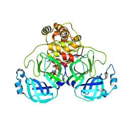
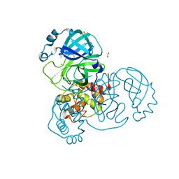
THE CRYSTAL STRUCTURE OF COVID-19 MAIN PROTEASE apo form at 2.2 angstrom
Descriptor: 3C-like proteinase
Authors:Liu, X, Zhao, Y, Yang, H, Rao, Z.
Deposit date:2021-02-21
Release date:2022-03-02
Last modified:2023-11-29
Method:X-RAY DIFFRACTION (2.19 Å)
Cite:Structural basis for replicase polyprotein cleavage and substrate specificity of main protease from SARS-CoV-2.
Proc.Natl.Acad.Sci.USA, 119, 2022
7NIJ
DownloadVisualize
BU of 7nij by Molmil
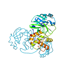
SARS-CoV-2 main protease (Mpro) in a novel conformational state.
Descriptor: 3C-like proteinase nsp5
Authors:Battistutta, R, Fornasier, E, Giachin, G.
Deposit date:2021-02-12
Release date:2022-03-02
Last modified:2024-01-31
Method:X-RAY DIFFRACTION (1.58 Å)
Cite:A new inactive conformation of SARS-CoV-2 main protease.
Acta Crystallogr D Struct Biol, 78, 2022
7NTQ
DownloadVisualize
BU of 7ntq by Molmil
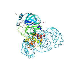
Crystal structure of the SARS-CoV-2 Main Protease complexed with N-(pyridin-3-ylmethyl)thioformamide
Descriptor: 3C-like proteinase, DIMETHYL SULFOXIDE, FORMIC ACID, ...
Authors:Dupre, E, Villeret, V, Hanoulle, X.
Deposit date:2021-03-10
Release date:2022-03-02
Last modified:2024-02-07
Method:X-RAY DIFFRACTION (1.495 Å)
Cite:Novel dithiocarbamates selectively inhibit 3CL protease of SARS-CoV-2 and other coronaviruses.
Eur.J.Med.Chem., 250, 2023
7NTT
DownloadVisualize
BU of 7ntt by Molmil
Crystal structure of the SARS-CoV-2 Main Protease
Descriptor: 3C-like proteinase, FORMIC ACID, SODIUM ION
Authors:Dupre, E, Villeret, V, Hanoulle, X.
Deposit date:2021-03-10
Release date:2022-03-02
Last modified:2024-01-31
Method:X-RAY DIFFRACTION (1.743 Å)
Cite:Crystal structure of the SARS-CoV-2 Main Protease
To Be Published
7NTW
DownloadVisualize
BU of 7ntw by Molmil
Crystal structure of the SARS-CoV-2 Main Protease with a Zinc ion coordinated in the active site
Descriptor: 3C-like proteinase, DIMETHYL SULFOXIDE, FORMIC ACID, ...
Authors:Dupre, E, Villeret, V, Hanoulle, X.
Deposit date:2021-03-11
Release date:2022-03-02
Last modified:2024-01-31
Method:X-RAY DIFFRACTION (1.815 Å)
Cite:Crystal structure of the SARS-CoV-2 Main Protease with a Zinc ion coordinated in the active site
To Be Published
7R10
DownloadVisualize
BU of 7r10 by Molmil
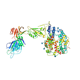
Dissociated S1 domain of Alpha Variant SARS-CoV-2 Spike bound to ACE2
Descriptor: 2-acetamido-2-deoxy-beta-D-glucopyranose, Angiotensin-converting enzyme 2, Spike glycoprotein, ...
Authors:Benton, D.J, Wrobel, A.G, Gamblin, S.J.
Deposit date:2022-02-02
Release date:2022-03-02
Last modified:2022-03-16
Method:ELECTRON MICROSCOPY (4 Å)
Cite:Evolution of the SARS-CoV-2 spike protein in the human host.
Nat Commun, 13, 2022

222415

건을2024-07-10부터공개중

PDB statisticsPDBj update infoContact PDBjnumon