 Movie
Movie Controller


+ Open data

- Basic information
| Entry | Database: PDB / ID: 1akn | ||||||
|---|---|---|---|---|---|---|---|
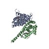
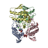
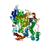
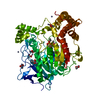
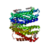
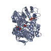
| Title | STRUCTURE OF BILE-SALT ACTIVATED LIPASE | ||||||
 Components Components | BILE-SALT ACTIVATED LIPASE | ||||||
 Keywords Keywords | HYDROLASE / SERINE ESTERASE / LIPID DEGRADATION / GLYCOPROTEIN / CARBOXYLIC ESTERASE / HYDROLASE() | ||||||
| Function / homology |  Function and homology information Function and homology informationretinyl-palmitate esterase activity / acetylesterase / ceramide catabolic process / sterol esterase / sterol ester esterase activity / pancreatic juice secretion / acetylesterase activity / triacylglycerol lipase / triacylglycerol lipase activity / extracellular region / cytoplasm Similarity search - Function | ||||||
| Biological species |  | ||||||
| Method |  X-RAY DIFFRACTION / MOLECULAR REPLACEMENT / Resolution: 2.8 Å X-RAY DIFFRACTION / MOLECULAR REPLACEMENT / Resolution: 2.8 Å | ||||||
 Authors Authors | Wang, X. / Zhang, X. | ||||||
 Citation Citation | Journal: Structure / Year: 1997 Title: The crystal structure of bovine bile salt activated lipase: insights into the bile salt activation mechanism. Authors: Wang, X. / Wang, C.S. / Tang, J. / Dyda, F. / Zhang, X.C. | ||||||
| History |
|
- Structure visualization
| Structure viewer | Molecule: MolmilJmol/JSmol |
|---|
- Downloads & links
-Download
| PDBx/mmCIF format | 1akn.cif.gz | 113.1 KB | Display | PDBx/mmCIF format |
|---|---|---|---|---|
| PDB format | pdb1akn.ent.gz | 90.4 KB | Display | PDB format |
| PDBx/mmJSON format | 1akn.json.gz | Tree view | PDBx/mmJSON format | |
| Others |  Other downloads Other downloads |
-Validation report
| Summary document | 1akn_validation.pdf.gz | 389.6 KB | Display | wwPDB validaton report |
|---|---|---|---|---|
| Full document | 1akn_full_validation.pdf.gz | 404.2 KB | Display | |
| Data in XML | 1akn_validation.xml.gz | 13.6 KB | Display | |
| Data in CIF | 1akn_validation.cif.gz | 19.8 KB | Display | |
| Arichive directory | https://data.pdbj.org/pub/pdb/validation_reports/ak/1aknftp://data.pdbj.org/pub/pdb/validation_reports/ak/1akn | HTTPS FTP |
-Related structure data
| Related structure data |  1aqlC  1ace S: Starting model for refinement C: citing same article ( |
|---|---|
| Similar structure data |

-Links
 PDBj
PDBj
- Assembly
| Deposited unit | 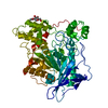
| ||||||||
|---|---|---|---|---|---|---|---|---|---|
| 1 |
| ||||||||
| Unit cell |
|
-Components
| #1: Protein | Mass: 63611.363 Da / Num. of mol.: 1 / Source method: isolated from a natural source / Source: (natural) |
|---|---|
| #2: Sugar | ChemComp-NAG /   Type: D-saccharide, beta linking / Mass: 221.208 Da / Num. of mol.: 1 Type: D-saccharide, beta linking / Mass: 221.208 Da / Num. of mol.: 1Source method: isolated from a genetically manipulated source Formula: C8H15NO6 |
-Experimental details
-Experiment
| Experiment | Method: X-RAY DIFFRACTION / Number of used crystals: 1 |
|---|
- Sample preparation
| Crystal | Density Matthews: 2.93 Å3/Da / Density % sol: 58 % | ||||||||||||||||||||||||||||||||||||||||
|---|---|---|---|---|---|---|---|---|---|---|---|---|---|---|---|---|---|---|---|---|---|---|---|---|---|---|---|---|---|---|---|---|---|---|---|---|---|---|---|---|---|
| Crystal grow | pH: 7 / Details: pH 7. | ||||||||||||||||||||||||||||||||||||||||
| Crystal grow | *PLUS Temperature: 20 ℃ / Method: vapor diffusion, hanging drop | ||||||||||||||||||||||||||||||||||||||||
| Components of the solutions | *PLUS
|
-Data collection
| Diffraction | Mean temperature: 288 K |
|---|---|
| Diffraction source | Source: ROTATING ANODE / Type: RIGAKU RUH2R / Wavelength: 1.5418 |
| Detector | Type: RIGAKU RAXIS II / Detector: IMAGE PLATE / Date: Dec 1, 1996 / Details: MIRROR |
| Radiation | Monochromator: NI FILTER / Monochromatic (M) / Laue (L): M / Scattering type: x-ray |
| Radiation wavelength | Wavelength: 1.5418 Å / Relative weight: 1 |
| Reflection | Resolution: 2.8→33.7 Å / Num. obs: 18614 / % possible obs: 98.4 % / Observed criterion σ(I): 0 / Redundancy: 2.53 % / Biso Wilson estimate: 66.1 Å2 / Rmerge(I) obs: 0.066 / Net I/σ(I): 13.8 |
| Reflection shell | Resolution: 2.8→2.9 Å / Redundancy: 1.3 % / Rmerge(I) obs: 0.39 / Mean I/σ(I) obs: 2.8 / % possible all: 91.2 |
| Reflection | *PLUS Lowest resolution: 34 Å |
| Reflection shell | *PLUS % possible obs: 99.8 % / Rmerge(I) obs: 0.391 |
- Processing
| Software |
| ||||||||||||||||||||||||||||||||||||||||||||||||||||||||||||
|---|---|---|---|---|---|---|---|---|---|---|---|---|---|---|---|---|---|---|---|---|---|---|---|---|---|---|---|---|---|---|---|---|---|---|---|---|---|---|---|---|---|---|---|---|---|---|---|---|---|---|---|---|---|---|---|---|---|---|---|---|---|
| Refinement | Method to determine structure: MOLECULAR REPLACEMENT Starting model: PDB ENTRY 1ACE 1ace Resolution: 2.8→8 Å Details: AT FINAL STEP, ALL DATA IN 8 - 2.8 A WERE USED TO CARRY OUT A RESTRAINED TEMPERATURE FACTOR REFINEMENT BY TNT.
| ||||||||||||||||||||||||||||||||||||||||||||||||||||||||||||
| Displacement parameters | Biso mean: 47.6 Å2 | ||||||||||||||||||||||||||||||||||||||||||||||||||||||||||||
| Refinement step | Cycle: LAST / Resolution: 2.8→8 Å
| ||||||||||||||||||||||||||||||||||||||||||||||||||||||||||||
| Refine LS restraints |
| ||||||||||||||||||||||||||||||||||||||||||||||||||||||||||||
| Xplor file |
| ||||||||||||||||||||||||||||||||||||||||||||||||||||||||||||
| Software | *PLUS Name: X-PLOR / Version: 3.1 / Classification: refinement | ||||||||||||||||||||||||||||||||||||||||||||||||||||||||||||
| Refinement | *PLUS Rfactor obs: 0.216 / Rfactor Rfree: 0.283 | ||||||||||||||||||||||||||||||||||||||||||||||||||||||||||||
| Solvent computation | *PLUS | ||||||||||||||||||||||||||||||||||||||||||||||||||||||||||||
| Displacement parameters | *PLUS | ||||||||||||||||||||||||||||||||||||||||||||||||||||||||||||
| Refine LS restraints | *PLUS
|
