 Movie
Movie Controller

[English] 日本語
 Yorodumi
- PDB-1aql: CRYSTAL STRUCTURE OF BOVINE BILE-SALT ACTIVATED LIPASE COMPLEXED ... -
+ Open data

- Basic information
| Entry | Database: PDB / ID: 1aql | ||||||
|---|---|---|---|---|---|---|---|
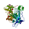
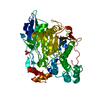
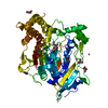
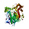
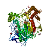
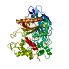
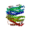
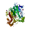
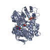
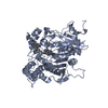
| Title | CRYSTAL STRUCTURE OF BOVINE BILE-SALT ACTIVATED LIPASE COMPLEXED WITH TAUROCHOLATE | ||||||
 Components Components | BILE-SALT ACTIVATED LIPASE | ||||||
 Keywords Keywords | HYDROLASE / SERINE ESTERASE / LIPID DEGRADATION / GLYCOPROTEIN | ||||||
| Function / homology |  Function and homology information Function and homology informationretinyl-palmitate esterase activity / acetylesterase / ceramide catabolic process / sterol esterase / sterol ester esterase activity / pancreatic juice secretion / acetylesterase activity / triacylglycerol lipase / triacylglycerol lipase activity / extracellular region ...retinyl-palmitate esterase activity / acetylesterase / ceramide catabolic process / sterol esterase / sterol ester esterase activity / pancreatic juice secretion / acetylesterase activity / triacylglycerol lipase / triacylglycerol lipase activity / extracellular region / membrane / cytoplasm Similarity search - Function | ||||||
| Biological species |  | ||||||
| Method |  X-RAY DIFFRACTION / MOLECULAR REPLACEMENT / Resolution: 2.8 Å X-RAY DIFFRACTION / MOLECULAR REPLACEMENT / Resolution: 2.8 Å | ||||||
 Authors Authors | Wang, X. / Zhang, X. | ||||||
 Citation Citation | Journal: Structure / Year: 1997 Title: The crystal structure of bovine bile salt activated lipase: insights into the bile salt activation mechanism. Authors: Wang, X. / Wang, C.S. / Tang, J. / Dyda, F. / Zhang, X.C. | ||||||
| History |
|
- Structure visualization
| Structure viewer | Molecule: MolmilJmol/JSmol |
|---|
- Downloads & links
-Download
| PDBx/mmCIF format | 1aql.cif.gz | 214.9 KB | Display | PDBx/mmCIF format |
|---|---|---|---|---|
| PDB format | pdb1aql.ent.gz | 174.3 KB | Display | PDB format |
| PDBx/mmJSON format | 1aql.json.gz | Tree view | PDBx/mmJSON format | |
| Others |  Other downloads Other downloads |
-Validation report
| Arichive directory | https://data.pdbj.org/pub/pdb/validation_reports/aq/1aqlftp://data.pdbj.org/pub/pdb/validation_reports/aq/1aql | HTTPS FTP |
|---|
-Related structure data
| Related structure data |  1aknSC S: Starting model for refinement C: citing same article ( |
|---|---|
| Similar structure data |
-Links
 PDBj
PDBj
- Assembly
| Deposited unit | 
| ||||||||
|---|---|---|---|---|---|---|---|---|---|
| 1 | 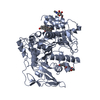
| ||||||||
| 2 | 
| ||||||||
| Unit cell |
| ||||||||
| Noncrystallographic symmetry (NCS) | NCS oper: (Code: given Matrix: (0.352585, 0.46694, 0.810957), Vector: |
-Components
| #1: Protein | Mass: 59065.461 Da / Num. of mol.: 2 / Source method: isolated from a natural source / Source: (natural) #2: Sugar |   Type: D-saccharide, beta linking / Mass: 221.208 Da / Num. of mol.: 2 Type: D-saccharide, beta linking / Mass: 221.208 Da / Num. of mol.: 2Source method: isolated from a genetically manipulated source Formula: C8H15NO6 #3: Chemical | ChemComp-TCH /   Mass: 515.703 Da / Num. of mol.: 4 / Source method: obtained synthetically / Formula: C26H45NO7S Mass: 515.703 Da / Num. of mol.: 4 / Source method: obtained synthetically / Formula: C26H45NO7SHas protein modification | Y | |
|---|
-Experimental details
-Experiment
| Experiment | Method: X-RAY DIFFRACTION / Number of used crystals: 1 |
|---|
- Sample preparation
| Crystal | Density Matthews: 3.2 Å3/Da / Density % sol: 61.5 % | ||||||||||||||||||||||||||||||||||||||||||||||||||
|---|---|---|---|---|---|---|---|---|---|---|---|---|---|---|---|---|---|---|---|---|---|---|---|---|---|---|---|---|---|---|---|---|---|---|---|---|---|---|---|---|---|---|---|---|---|---|---|---|---|---|---|
| Crystal grow | pH: 7 / Details: pH 7 | ||||||||||||||||||||||||||||||||||||||||||||||||||
| Crystal grow | *PLUS Temperature: 20 ℃ / Method: vapor diffusion, hanging drop | ||||||||||||||||||||||||||||||||||||||||||||||||||
| Components of the solutions | *PLUS
|
-Data collection
| Diffraction | Mean temperature: 293 K |
|---|---|
| Diffraction source | Source: ROTATING ANODE / Type: SIEMENS / Wavelength: 1.5418 |
| Detector | Type: SIEMENS / Detector: AREA DETECTOR / Date: Mar 1, 1997 |
| Radiation | Monochromator: CRYSTAL / Monochromatic (M) / Laue (L): M / Scattering type: x-ray |
| Radiation wavelength | Wavelength: 1.5418 Å / Relative weight: 1 |
| Reflection | Resolution: 2.8→17 Å / Num. obs: 33877 / % possible obs: 82.7 % / Observed criterion σ(I): 0.5 / Redundancy: 1.8 % / Biso Wilson estimate: 43.5 Å2 / Rsym value: 0.117 / Net I/σ(I): 9.1 |
| Reflection shell | Resolution: 2.8→2.9 Å / Redundancy: 1.3 % / Mean I/σ(I) obs: 2.4 / Rsym value: 0.206 / % possible all: 48.3 |
| Reflection | *PLUS Rmerge(I) obs: 0.117 |
| Reflection shell | *PLUS % possible obs: 48.3 % / Rmerge(I) obs: 0.206 |
- Processing
| Software |
| ||||||||||||||||||||||||||||||||||||||||||||||||||||||||||||
|---|---|---|---|---|---|---|---|---|---|---|---|---|---|---|---|---|---|---|---|---|---|---|---|---|---|---|---|---|---|---|---|---|---|---|---|---|---|---|---|---|---|---|---|---|---|---|---|---|---|---|---|---|---|---|---|---|---|---|---|---|---|
| Refinement | Method to determine structure: MOLECULAR REPLACEMENT Starting model: PDB ENTRY 1AKN Resolution: 2.8→8 Å / σ(F): 1.5 Details: AT FINAL STEP, A RESTRAINED TEMPERATURE FACTOR REFINEMENT WAS CARRIED OUT BY TNT.
| ||||||||||||||||||||||||||||||||||||||||||||||||||||||||||||
| Displacement parameters | Biso mean: 43.7 Å2
| ||||||||||||||||||||||||||||||||||||||||||||||||||||||||||||
| Refinement step | Cycle: LAST / Resolution: 2.8→8 Å
| ||||||||||||||||||||||||||||||||||||||||||||||||||||||||||||
| Refine LS restraints |
| ||||||||||||||||||||||||||||||||||||||||||||||||||||||||||||
| Refine LS restraints NCS | NCS model details: RESTRAINTS | ||||||||||||||||||||||||||||||||||||||||||||||||||||||||||||
| Xplor file |
| ||||||||||||||||||||||||||||||||||||||||||||||||||||||||||||
| Software | *PLUS Name: X-PLOR / Version: 3.1 / Classification: refinement | ||||||||||||||||||||||||||||||||||||||||||||||||||||||||||||
| Refine LS restraints | *PLUS
|
