 Movie
Movie Controller

[English] 日本語
 Yorodumi
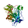
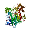
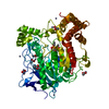
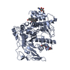
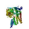
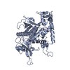
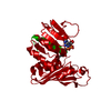
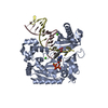
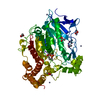
- PDB-6h0v: Crystal structure of tabun surrogate NEDPA inhibited recombinant ... -
+ Open data

- Basic information
| Entry | Database: PDB / ID: 6h0v | |||||||||
|---|---|---|---|---|---|---|---|---|---|---|
| Title | Crystal structure of tabun surrogate NEDPA inhibited recombinant human bile salt activated lipase | |||||||||
 Components Components | Bile salt-activated lipase | |||||||||
 Keywords Keywords | HYDROLASE / Lipase / alpha-beta hydrolase / tabun inhibition | |||||||||
| Function / homology |  Function and homology information Function and homology informationretinyl-palmitate esterase activity / Digestion of dietary lipid / acetylesterase / ceramide catabolic process / sterol esterase / sterol ester esterase activity / pancreatic juice secretion / acetylesterase activity / intestinal cholesterol absorption / Developmental Lineage of Pancreatic Acinar Cells ...retinyl-palmitate esterase activity / Digestion of dietary lipid / acetylesterase / ceramide catabolic process / sterol esterase / sterol ester esterase activity / pancreatic juice secretion / acetylesterase activity / intestinal cholesterol absorption / Developmental Lineage of Pancreatic Acinar Cells / triacylglycerol lipase / triacylglycerol lipase activity / catalytic activity / lipid metabolic process / heparin binding / hydrolase activity / : / extracellular exosome / extracellular region / membrane / cytoplasm Similarity search - Function | |||||||||
| Biological species |  Homo sapiens (human) Homo sapiens (human) | |||||||||
| Method |  X-RAY DIFFRACTION / SYNCHROTRON / MOLECULAR REPLACEMENT / Resolution: 2.2 Å X-RAY DIFFRACTION / SYNCHROTRON / MOLECULAR REPLACEMENT / Resolution: 2.2 Å | |||||||||
 Authors Authors | Touvrey, C. / Brazzolotto, X. / Nachon, F. | |||||||||
| Funding support |  France, 2items France, 2items
| |||||||||
 Citation Citation | Journal: Toxicology / Year: 2019 Title: X-ray structures of human bile-salt activated lipase conjugated to nerve agents surrogates. Authors: Touvrey, C. / Courageux, C. / Guillon, V. / Terreux, R. / Nachon, F. / Brazzolotto, X. | |||||||||
| History |
|
- Structure visualization
| Structure viewer | Molecule: MolmilJmol/JSmol |
|---|
- Downloads & links
-Download
| PDBx/mmCIF format | 6h0v.cif.gz | 144.9 KB | Display | PDBx/mmCIF format |
|---|---|---|---|---|
| PDB format | pdb6h0v.ent.gz | 89.1 KB | Display | PDB format |
| PDBx/mmJSON format | 6h0v.json.gz | Tree view | PDBx/mmJSON format | |
| Others |  Other downloads Other downloads |
-Validation report
| Arichive directory | https://data.pdbj.org/pub/pdb/validation_reports/h0/6h0vftp://data.pdbj.org/pub/pdb/validation_reports/h0/6h0v | HTTPS FTP |
|---|
-Related structure data
| Related structure data | 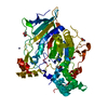 6h0tC  6h18C  6h19C  6h1aC  1f6wS S: Starting model for refinement C: citing same article ( |
|---|---|
| Similar structure data |

-Links
 PDBj
PDBj
- Assembly
| Deposited unit | 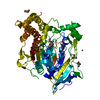
| ||||||||||||
|---|---|---|---|---|---|---|---|---|---|---|---|---|---|
| 1 |
| ||||||||||||
| Unit cell |
|
-Components
| #1: Protein | Mass: 60877.816 Da / Num. of mol.: 1 / Mutation: N186D, A298D, 534STOP Source method: isolated from a genetically manipulated source Details: SUN = serine residue inhibited by tabun (already in PDB database please see 2JEZ) Source: (gene. exp.) Homo sapiens (human) / Gene: CEL, BAL / Production host:   Cricetulus griseus (Chinese hamster) Cricetulus griseus (Chinese hamster)References: UniProt: P19835, sterol esterase, triacylglycerol lipase | ||||
|---|---|---|---|---|---|
| #2: Chemical | ChemComp-ZN /   Mass: 65.409 Da / Num. of mol.: 5 / Source method: obtained synthetically / Formula: Zn Mass: 65.409 Da / Num. of mol.: 5 / Source method: obtained synthetically / Formula: Zn#3: Chemical | ChemComp-ACT /   Mass: 59.044 Da / Num. of mol.: 4 / Source method: obtained synthetically / Formula: C2H3O2 Mass: 59.044 Da / Num. of mol.: 4 / Source method: obtained synthetically / Formula: C2H3O2#4: Water | ChemComp-HOH / |  Mass: 18.015 Da / Num. of mol.: 74 / Source method: isolated from a natural source / Formula: H2O Mass: 18.015 Da / Num. of mol.: 74 / Source method: isolated from a natural source / Formula: H2O |
-Experimental details
-Experiment
| Experiment | Method: X-RAY DIFFRACTION / Number of used crystals: 1 |
|---|
- Sample preparation
| Crystal | Density Matthews: 2.55 Å3/Da / Density % sol: 51.83 % |
|---|---|
| Crystal grow | Temperature: 293 K / Method: vapor diffusion, hanging drop / Details: PEG 8000, cacodylate, Zinc acetate |
-Data collection
| Diffraction | Mean temperature: 100 K |
|---|---|
| Diffraction source | Source: SYNCHROTRON / Site: ESRF / Beamline: MASSIF-1 / Wavelength: 0.966 Å |
| Detector | Type: DECTRIS PILATUS3 2M / Detector: PIXEL / Date: Jul 20, 2016 |
| Radiation | Protocol: SINGLE WAVELENGTH / Monochromatic (M) / Laue (L): M / Scattering type: x-ray |
| Radiation wavelength | Wavelength: 0.966 Å / Relative weight: 1 |
| Reflection | Resolution: 2.2→48.11 Å / Num. obs: 28933 / % possible obs: 94.42 % / Redundancy: 4 % / Biso Wilson estimate: 33.11 Å2 / Net I/σ(I): 13.52 |
| Reflection shell | Resolution: 2.2→2.279 Å / Redundancy: 4.1 % / Mean I/σ(I) obs: 5.21 / Num. unique obs: 2882 / % possible all: 95.15 |
- Processing
| Software |
| |||||||||||||||||||||||||||||||||||||||||||||||||||||||||||||||
|---|---|---|---|---|---|---|---|---|---|---|---|---|---|---|---|---|---|---|---|---|---|---|---|---|---|---|---|---|---|---|---|---|---|---|---|---|---|---|---|---|---|---|---|---|---|---|---|---|---|---|---|---|---|---|---|---|---|---|---|---|---|---|---|---|
| Refinement | Method to determine structure: MOLECULAR REPLACEMENT Starting model: 1f6w Resolution: 2.2→48.11 Å / SU ML: 0.2748 / Cross valid method: FREE R-VALUE / σ(F): 1.36 / Phase error: 28.0005
| |||||||||||||||||||||||||||||||||||||||||||||||||||||||||||||||
| Solvent computation | Shrinkage radii: 0.9 Å / VDW probe radii: 1.11 Å | |||||||||||||||||||||||||||||||||||||||||||||||||||||||||||||||
| Displacement parameters | Biso mean: 42.27 Å2 | |||||||||||||||||||||||||||||||||||||||||||||||||||||||||||||||
| Refinement step | Cycle: LAST / Resolution: 2.2→48.11 Å
| |||||||||||||||||||||||||||||||||||||||||||||||||||||||||||||||
| Refine LS restraints |
| |||||||||||||||||||||||||||||||||||||||||||||||||||||||||||||||
| LS refinement shell |
|
