National Institutes of Health/National Institute Of Allergy and Infectious Diseases (NIH/NIAID)
R01 AI092571
United States
National Institutes of Health/National Institute Of Allergy and Infectious Diseases (NIH/NIAID)
GM080533
United States
Medical Research Council (MRC, United Kingdom)
MC_UU_12014/10
United Kingdom
Citation
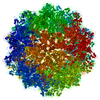
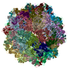
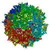
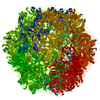
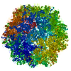
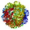
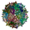
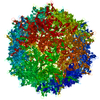
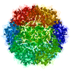
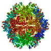
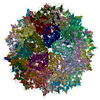
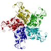
Journal: J Virol / Year: 2019 Title: Examination and Reconstruction of Three Ancient Endogenous Parvovirus Capsid Protein Gene Remnants Found in Rodent Genomes. Authors: Heather M Callaway / Suriyasri Subramanian / Christian A Urbina / Karen N Barnard / Robert A Dick / Carol M Bator / Susan L Hafenstein / Robert J Gifford / Colin R Parrish / Abstract: Parvovirus-derived endogenous viral elements (EVEs) have been found in the genomes of many different animal species, resulting from integration events that may have occurred from more than 50 million ...Parvovirus-derived endogenous viral elements (EVEs) have been found in the genomes of many different animal species, resulting from integration events that may have occurred from more than 50 million years ago to much more recently. Here, we further investigate the properties of autonomous parvovirus EVEs and describe their relationships to contemporary viruses. While we did not find any intact capsid protein open reading frames in the integrated viral sequences, we examined three EVEs that were repaired to form full-length sequences with relatively few changes. These sequences were found in the genomes of (brown rat), (Algerian mouse), and (wood mouse). The sequence was not present in the genomes of the closely related species , , , and , indicating that it was less than 2 million years old, and the and sequences were not found in the published genomes of other mouse species, also indicating relatively recent insertions. The VP2 sequence assembled into capsids, which had high thermal stability, bound the sialic acid -acetylneuraminic acid, and entered murine L cells. The 3.89-Å structure of the virus-like particles (VLPs), determined using cryo-electron microscopy, showed similarities to rodent and porcine parvovirus capsids. The repaired VP2 sequences from and did not assemble as first prepared, but chimeras combining capsid surface loops from with canine parvovirus assembled, allowing some of that capsid's structures and functions to be examined. Parvovirus endogenous viral elements (EVEs) that have been incorporated into the genomes of different animals represent remnants of the DNA sequences of ancient viruses that infected the ancestors of those animals millions of years ago, but we know little about their properties or how they differ from currently circulating parvoviruses. By expressing the capsid proteins of different parvovirus EVEs that were found integrated into the genomes of three different rodents, we can examine their structures and functions. A VP2 (major capsid protein) EVE sequence from a mouse genome assembled into capsids that had a similar structure and biophysical properties to extant parvoviruses and also bound sialic acids and entered rodent cells. Chimeras formed from combinations of canine parvovirus and portions of the parvovirus sequences from the brown rat genome allowed us to examine the structures and functions of the surface loops of that EVE capsid.
In the structure databanks used in Yorodumi, some data are registered as the other names, "COVID-19 virus" and "2019-nCoV". Here are the details of the virus and the list of structure data.
Jan 31, 2019. EMDB accession codes are about to change! (news from PDBe EMDB page)
EMDB accession codes are about to change! (news from PDBe EMDB page)
The allocation of 4 digits for EMDB accession codes will soon come to an end. Whilst these codes will remain in use, new EMDB accession codes will include an additional digit and will expand incrementally as the available range of codes is exhausted. The current 4-digit format prefixed with “EMD-” (i.e. EMD-XXXX) will advance to a 5-digit format (i.e. EMD-XXXXX), and so on. It is currently estimated that the 4-digit codes will be depleted around Spring 2019, at which point the 5-digit format will come into force.
The EM Navigator/Yorodumi systems omit the EMD- prefix.
Related info.:Q: What is EMD? / ID/Accession-code notation in Yorodumi/EM Navigator
Yorodumi is a browser for structure data from EMDB, PDB, SASBDB, etc.
This page is also the successor to EM Navigator detail page, and also detail information page/front-end page for Omokage search.
The word "yorodu" (or yorozu) is an old Japanese word meaning "ten thousand". "mi" (miru) is to see.
Related info.:EMDB / PDB / SASBDB / Comparison of 3 databanks / Yorodumi Search / Aug 31, 2016. New EM Navigator & Yorodumi / Yorodumi Papers / Jmol/JSmol / Function and homology information / Changes in new EM Navigator and Yorodumi
 Movie
Movie Controller

 Yorodumi
 Open data
 Basic information
 Components
 Keywords
 Function and homology information
 Authors
 United States,
  United Kingdom, 3items
  Citation
 Structure visualization
 Downloads & links
 Other downloads
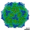


 PDBj
PDBj Assembly


 Trichoplusia ni (cabbage looper)
 Sample preparation
 Parvoviridae (virus) / Strain: Mus spretus endogenous viral element
 Electron microscopy imaging
 FIELD EMISSION GUN / Accelerating voltage: 300 kV / Illumination mode: FLOOD BEAM
 Processing