 Movie
Movie Controller

[English] 日本語
 Yorodumi
- PDB-5uvl: Serial Millisecond Crystallography of Membrane and Soluble Protei... -
+ Open data

- Basic information
| Entry | Database: PDB / ID: 5uvl | |||||||||||||||||||||||||||
|---|---|---|---|---|---|---|---|---|---|---|---|---|---|---|---|---|---|---|---|---|---|---|---|---|---|---|---|---|
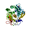
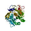
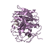
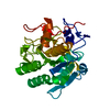
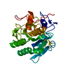
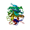
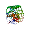
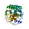
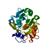
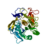
| Title | Serial Millisecond Crystallography of Membrane and Soluble Protein Micro-crystals using Synchrotron Radiation | |||||||||||||||||||||||||||
 Components Components | Proteinase K | |||||||||||||||||||||||||||
 Keywords Keywords | HYDROLASE | |||||||||||||||||||||||||||
| Function / homology |  Function and homology information Function and homology informationpeptidase K / serine-type endopeptidase activity / proteolysis / extracellular region / metal ion bindingSimilarity search - Function | |||||||||||||||||||||||||||
| Biological species |  Parengyodontium album (fungus) Parengyodontium album (fungus) | |||||||||||||||||||||||||||
| Method | X-RAY DIFFRACTION / SYNCHROTRON / MOLECULAR REPLACEMENT / Resolution: 2.65 Å | |||||||||||||||||||||||||||
 Authors Authors | Martin-Garcia, J.M. / Conrad, C.E. / Nelson, G. / Stander, N. / Zatsepin, N.A. / Zook, J. / Zhu, L. / Geiger, J. / Chun, E. / Kissick, D. ...Martin-Garcia, J.M. / Conrad, C.E. / Nelson, G. / Stander, N. / Zatsepin, N.A. / Zook, J. / Zhu, L. / Geiger, J. / Chun, E. / Kissick, D. / Hilgart, M.C. / Ogata, C. / Ishchenko, A. / Nagaratnam, N. / Roy-Chowdhury, S. / Coe, J. / Subramanian, G. / Schaffer, A. / James, D. / Ketawala, G. / Venugopalan, N. / Xu, S. / Corcoran, S. / Ferguson, D. / Weierstall, U. / Spence, J.C.H. / Cherezov, V. / Fromme, P. / Fischetti, R.F. / Liu, W. | |||||||||||||||||||||||||||
| Funding support |  United States, 8items United States, 8items
| |||||||||||||||||||||||||||
 Citation Citation | Journal: IUCrJ / Year: 2017 Title: Serial millisecond crystallography of membrane and soluble protein microcrystals using synchrotron radiation. Authors: Martin-Garcia, J.M. / Conrad, C.E. / Nelson, G. / Stander, N. / Zatsepin, N.A. / Zook, J. / Zhu, L. / Geiger, J. / Chun, E. / Kissick, D. / Hilgart, M.C. / Ogata, C. / Ishchenko, A. / ...Authors: Martin-Garcia, J.M. / Conrad, C.E. / Nelson, G. / Stander, N. / Zatsepin, N.A. / Zook, J. / Zhu, L. / Geiger, J. / Chun, E. / Kissick, D. / Hilgart, M.C. / Ogata, C. / Ishchenko, A. / Nagaratnam, N. / Roy-Chowdhury, S. / Coe, J. / Subramanian, G. / Schaffer, A. / James, D. / Ketwala, G. / Venugopalan, N. / Xu, S. / Corcoran, S. / Ferguson, D. / Weierstall, U. / Spence, J.C.H. / Cherezov, V. / Fromme, P. / Fischetti, R.F. / Liu, W. | |||||||||||||||||||||||||||
| History |
|
- Structure visualization
| Structure viewer | Molecule: MolmilJmol/JSmol |
|---|
- Downloads & links
-Download
| PDBx/mmCIF format | 5uvl.cif.gz | 65.5 KB | Display | PDBx/mmCIF format |
|---|---|---|---|---|
| PDB format | pdb5uvl.ent.gz | 46.6 KB | Display | PDB format |
| PDBx/mmJSON format | 5uvl.json.gz | Tree view | PDBx/mmJSON format | |
| Others |  Other downloads Other downloads |
-Validation report
| Arichive directory | https://data.pdbj.org/pub/pdb/validation_reports/uv/5uvlftp://data.pdbj.org/pub/pdb/validation_reports/uv/5uvl | HTTPS FTP |
|---|
-Related structure data
| Related structure data |  5uviC  5uvjC  5uvkC  5avjS C: citing same article ( S: Starting model for refinement |
|---|---|
| Similar structure data |


-Links
 PDBj
PDBj

- Assembly
| Deposited unit | 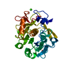
| ||||||||
|---|---|---|---|---|---|---|---|---|---|
| 1 |
| ||||||||
| Unit cell |
|
-Components
| #1: Protein | / Endopeptidase K / Tritirachium alkaline proteinase Mass: 28958.791 Da / Num. of mol.: 1 Source method: isolated from a genetically manipulated source Source: (gene. exp.) Parengyodontium album (fungus) / Gene: PROK / Production host: unidentified (others) / References: UniProt: P06873, peptidase K | ||
|---|---|---|---|
| #2: Chemical | Nitrate  Mass: 62.005 Da / Num. of mol.: 2 / Source method: obtained synthetically / Formula: NO3 Mass: 62.005 Da / Num. of mol.: 2 / Source method: obtained synthetically / Formula: NO3#3: Chemical |   Mass: 40.078 Da / Num. of mol.: 2 / Source method: obtained synthetically / Formula: Ca Mass: 40.078 Da / Num. of mol.: 2 / Source method: obtained synthetically / Formula: Ca |
-Experimental details
-Experiment
| Experiment | Method: X-RAY DIFFRACTION / Number of used crystals: 1 |
|---|
- Sample preparation
| Crystal | Density Matthews: 2.01 Å3/Da / Density % sol: 43.54 % |
|---|---|
| Crystal grow | Temperature: 293 K / Method: batch mode / pH: 6.5 Details: 0.1 M MES pH 6.5, 0.5 M sodium nitrate, 0.1 M calcium chloride |
-Data collection
| Diffraction | Mean temperature: 298 K |
|---|---|
| Diffraction source | Source: SYNCHROTRON / Site: APS / Beamline: 23-ID-D / Wavelength: 1.03 Å |
| Detector | Type: DECTRIS PILATUS3 6M / Detector: PIXEL / Date: Aug 17, 2016 |
| Radiation | Protocol: SINGLE WAVELENGTH / Monochromatic (M) / Laue (L): M / Scattering type: x-ray |
| Radiation wavelength | Wavelength: 1.03 Å / Relative weight: 1 |
| Reflection | Resolution: 2.65→50.01 Å / Num. obs: 6769 / % possible obs: 99.2 % / Redundancy: 104.8 % / Net I/σ(I): 4.6 |
- Processing
| Software |
| ||||||||||||||||||||||||||||||||||||||||||||||||||||||||||||||||||||||||||||||||||||||||||||||||||||||||||||||||||||||||||||||||||||||||||||||||||||||||||||||||||||||||||||||||||||||
|---|---|---|---|---|---|---|---|---|---|---|---|---|---|---|---|---|---|---|---|---|---|---|---|---|---|---|---|---|---|---|---|---|---|---|---|---|---|---|---|---|---|---|---|---|---|---|---|---|---|---|---|---|---|---|---|---|---|---|---|---|---|---|---|---|---|---|---|---|---|---|---|---|---|---|---|---|---|---|---|---|---|---|---|---|---|---|---|---|---|---|---|---|---|---|---|---|---|---|---|---|---|---|---|---|---|---|---|---|---|---|---|---|---|---|---|---|---|---|---|---|---|---|---|---|---|---|---|---|---|---|---|---|---|---|---|---|---|---|---|---|---|---|---|---|---|---|---|---|---|---|---|---|---|---|---|---|---|---|---|---|---|---|---|---|---|---|---|---|---|---|---|---|---|---|---|---|---|---|---|---|---|---|---|
| Refinement | Method to determine structure: MOLECULAR REPLACEMENT Starting model: 5AVJ Resolution: 2.65→50.01 Å / Cor.coef. Fo:Fc: 0.934 / Cor.coef. Fo:Fc free: 0.916 / SU B: 24.593 / SU ML: 0.434 / Cross valid method: THROUGHOUT / ESU R Free: 0.372 / Stereochemistry target values: MAXIMUM LIKELIHOOD / Details: HYDROGENS HAVE BEEN ADDED IN THE RIDING POSITIONS
| ||||||||||||||||||||||||||||||||||||||||||||||||||||||||||||||||||||||||||||||||||||||||||||||||||||||||||||||||||||||||||||||||||||||||||||||||||||||||||||||||||||||||||||||||||||||
| Solvent computation | Ion probe radii: 0.8 Å / Shrinkage radii: 0.8 Å / VDW probe radii: 1.2 Å / Solvent model: MASK | ||||||||||||||||||||||||||||||||||||||||||||||||||||||||||||||||||||||||||||||||||||||||||||||||||||||||||||||||||||||||||||||||||||||||||||||||||||||||||||||||||||||||||||||||||||||
| Displacement parameters | Biso mean: 60.277 Å2
| ||||||||||||||||||||||||||||||||||||||||||||||||||||||||||||||||||||||||||||||||||||||||||||||||||||||||||||||||||||||||||||||||||||||||||||||||||||||||||||||||||||||||||||||||||||||
| Refinement step | Cycle: 1 / Resolution: 2.65→50.01 Å
| ||||||||||||||||||||||||||||||||||||||||||||||||||||||||||||||||||||||||||||||||||||||||||||||||||||||||||||||||||||||||||||||||||||||||||||||||||||||||||||||||||||||||||||||||||||||
| Refine LS restraints |
|