 Movie
Movie Controller

[English] 日本語
 Yorodumi
- PDB-5uvi: Serial Millisecond Crystallography of Membrane and Soluble Protei... -
+ Open data

- Basic information
| Entry | Database: PDB / ID: 5uvi | |||||||||||||||||||||||||||
|---|---|---|---|---|---|---|---|---|---|---|---|---|---|---|---|---|---|---|---|---|---|---|---|---|---|---|---|---|
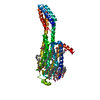
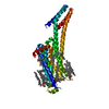
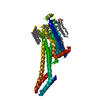
| Title | Serial Millisecond Crystallography of Membrane and Soluble Protein Micro-crystals using Synchrotron Radiation | |||||||||||||||||||||||||||
 Components Components | Adenosine receptor A2a,Soluble cytochrome b562,Adenosine receptor A2a | |||||||||||||||||||||||||||
 Keywords Keywords | MEMBRANE PROTEIN / GPCR | |||||||||||||||||||||||||||
| Function / homology |  Function and homology information Function and homology informationregulation of norepinephrine secretion / negative regulation of alpha-beta T cell activation / positive regulation of circadian sleep/wake cycle, sleep / positive regulation of acetylcholine secretion, neurotransmission / Adenosine P1 receptors / G protein-coupled adenosine receptor activity / response to purine-containing compound / G protein-coupled adenosine receptor signaling pathway / NGF-independant TRKA activation / Surfactant metabolism ...regulation of norepinephrine secretion / negative regulation of alpha-beta T cell activation / positive regulation of circadian sleep/wake cycle, sleep / positive regulation of acetylcholine secretion, neurotransmission / Adenosine P1 receptors / G protein-coupled adenosine receptor activity / response to purine-containing compound / G protein-coupled adenosine receptor signaling pathway / NGF-independant TRKA activation / Surfactant metabolism / synaptic transmission, dopaminergic / type 5 metabotropic glutamate receptor binding / negative regulation of vascular permeability / synaptic transmission, cholinergic / intermediate filament / presynaptic active zone / positive regulation of urine volume / response to caffeine / blood circulation / sensory perception / positive regulation of glutamate secretion / eating behavior / inhibitory postsynaptic potential / regulation of calcium ion transport / alpha-actinin binding / asymmetric synapse / axolemma / membrane depolarization / cellular defense response / prepulse inhibition / phagocytosis / positive regulation of synaptic transmission, glutamatergic / neuron projection morphogenesis / astrocyte activation / presynaptic modulation of chemical synaptic transmission / positive regulation of long-term synaptic potentiation / positive regulation of synaptic transmission, GABAergic / central nervous system development / positive regulation of protein secretion / response to amphetamine / regulation of mitochondrial membrane potential / positive regulation of apoptotic signaling pathway / apoptotic signaling pathway / synaptic transmission, glutamatergic / excitatory postsynaptic potential / locomotory behavior / electron transport chain / negative regulation of inflammatory response / vasodilation / adenylate cyclase-modulating G protein-coupled receptor signaling pathway / blood coagulation / cell-cell signaling / adenylate cyclase-activating G protein-coupled receptor signaling pathway / presynaptic membrane / G alpha (s) signalling events / phospholipase C-activating G protein-coupled receptor signaling pathway / negative regulation of neuron apoptotic process / calmodulin binding / periplasmic space / positive regulation of ERK1 and ERK2 cascade / electron transfer activity / postsynaptic membrane / iron ion binding / response to xenobiotic stimulus / inflammatory response / negative regulation of cell population proliferation / neuronal cell body / heme binding / apoptotic process / regulation of DNA-templated transcription / lipid binding / dendrite / protein-containing complex binding / glutamatergic synapse / enzyme binding / membrane / identical protein binding / plasma membrane Similarity search - Function | |||||||||||||||||||||||||||
| Biological species |  Homo sapiens (human) Homo sapiens (human) | |||||||||||||||||||||||||||
| Method |  X-RAY DIFFRACTION / SYNCHROTRON / MOLECULAR REPLACEMENT / Resolution: 3.2 Å X-RAY DIFFRACTION / SYNCHROTRON / MOLECULAR REPLACEMENT / Resolution: 3.2 Å | |||||||||||||||||||||||||||
 Authors Authors | Martin-Garcia, J.M. / Conrad, C.E. / Nelson, G. / Stander, N. / Zatsepin, N.A. / Zook, J. / Zhu, L. / Geiger, J. / Chun, E. / Kissick, D. ...Martin-Garcia, J.M. / Conrad, C.E. / Nelson, G. / Stander, N. / Zatsepin, N.A. / Zook, J. / Zhu, L. / Geiger, J. / Chun, E. / Kissick, D. / Hilgart, M.C. / Ogata, C. / Ishchenko, A. / Nagaratnam, N. / Roy-Chowdhury, S. / Coe, J. / Subramanian, G. / Schaffer, A. / James, D. / Ketawala, G. / Venugopalan, N. / Xu, S. / Corcoran, S. / Ferguson, D. / Weierstall, U. / Spence, J.C.H. / Cherezov, V. / Fromme, P. / Fischetti, R.F. / Liu, W. | |||||||||||||||||||||||||||
| Funding support |  United States, 8items United States, 8items
| |||||||||||||||||||||||||||
 Citation Citation | Journal: IUCrJ / Year: 2017 Title: Serial millisecond crystallography of membrane and soluble protein microcrystals using synchrotron radiation. Authors: Martin-Garcia, J.M. / Conrad, C.E. / Nelson, G. / Stander, N. / Zatsepin, N.A. / Zook, J. / Zhu, L. / Geiger, J. / Chun, E. / Kissick, D. / Hilgart, M.C. / Ogata, C. / Ishchenko, A. / ...Authors: Martin-Garcia, J.M. / Conrad, C.E. / Nelson, G. / Stander, N. / Zatsepin, N.A. / Zook, J. / Zhu, L. / Geiger, J. / Chun, E. / Kissick, D. / Hilgart, M.C. / Ogata, C. / Ishchenko, A. / Nagaratnam, N. / Roy-Chowdhury, S. / Coe, J. / Subramanian, G. / Schaffer, A. / James, D. / Ketwala, G. / Venugopalan, N. / Xu, S. / Corcoran, S. / Ferguson, D. / Weierstall, U. / Spence, J.C.H. / Cherezov, V. / Fromme, P. / Fischetti, R.F. / Liu, W. | |||||||||||||||||||||||||||
| History |
|
- Structure visualization
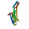
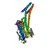
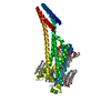
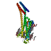
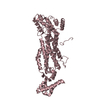
| Structure viewer | Molecule: MolmilJmol/JSmol |
|---|
- Downloads & links
-Download
| PDBx/mmCIF format | 5uvi.cif.gz | 98.1 KB | Display | PDBx/mmCIF format |
|---|---|---|---|---|
| PDB format | pdb5uvi.ent.gz | 70.8 KB | Display | PDB format |
| PDBx/mmJSON format | 5uvi.json.gz | Tree view | PDBx/mmJSON format | |
| Others |  Other downloads Other downloads |
-Validation report
| Arichive directory | https://data.pdbj.org/pub/pdb/validation_reports/uv/5uviftp://data.pdbj.org/pub/pdb/validation_reports/uv/5uvi | HTTPS FTP |
|---|
-Related structure data
| Related structure data |  5uvjC  5uvkC  5uvlC  5k2bS C: citing same article ( S: Starting model for refinement |
|---|---|
| Similar structure data |


-Links
 PDBj
PDBj


- Assembly
| Deposited unit | 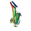
| ||||||||
|---|---|---|---|---|---|---|---|---|---|
| 1 |
| ||||||||
| Unit cell |
|
-Components
| #1: Protein | Mass: 49974.281 Da / Num. of mol.: 1 Source method: isolated from a genetically manipulated source Source: (gene. exp.) Homo sapiens (human), (gene. exp.) Gene: ADORA2A, ADORA2, cybC / Production host:   Spodoptera frugiperda (fall armyworm) / References: UniProt: P29274, UniProt: P0ABE7 Spodoptera frugiperda (fall armyworm) / References: UniProt: P29274, UniProt: P0ABE7 | ||||||
|---|---|---|---|---|---|---|---|
| #2: Chemical | ChemComp-ZMA / 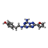  Mass: 337.336 Da / Num. of mol.: 1 / Source method: obtained synthetically / Formula: C16H15N7O2 / Comment: antagonist*YM Mass: 337.336 Da / Num. of mol.: 1 / Source method: obtained synthetically / Formula: C16H15N7O2 / Comment: antagonist*YM | ||||||
| #3: Chemical |   Mass: 386.654 Da / Num. of mol.: 3 / Source method: obtained synthetically / Formula: C27H46O Mass: 386.654 Da / Num. of mol.: 3 / Source method: obtained synthetically / Formula: C27H46O#4: Chemical |   Mass: 356.540 Da / Num. of mol.: 2 / Source method: obtained synthetically / Formula: C21H40O4 Mass: 356.540 Da / Num. of mol.: 2 / Source method: obtained synthetically / Formula: C21H40O4#5: Chemical | ChemComp-OLA / |   Mass: 282.461 Da / Num. of mol.: 1 / Source method: obtained synthetically / Formula: C18H34O2 Mass: 282.461 Da / Num. of mol.: 1 / Source method: obtained synthetically / Formula: C18H34O2Has protein modification | Y | |
-Experimental details
-Experiment
| Experiment | Method: X-RAY DIFFRACTION / Number of used crystals: 1 |
|---|
- Sample preparation
| Crystal | Density Matthews: 2.59 Å3/Da / Density % sol: 52.54 % |
|---|---|
| Crystal grow | Temperature: 293 K / Method: lipidic cubic phase / pH: 5 Details: 0.1 M sodium citrate pH 5.0, 32 % PEG 400, 75 mM sodium thocyanate |
-Data collection
| Diffraction | Mean temperature: 298 K |
|---|---|
| Diffraction source | Source: SYNCHROTRON / Site: APS / Beamline: 23-ID-D / Wavelength: 1.03 Å |
| Detector | Type: DECTRIS PILATUS3 6M / Detector: PIXEL / Date: Aug 16, 2016 |
| Radiation | Protocol: SINGLE WAVELENGTH / Monochromatic (M) / Laue (L): M / Scattering type: x-ray |
| Radiation wavelength | Wavelength: 1.03 Å / Relative weight: 1 |
| Reflection | Resolution: 3.2→45 Å / Num. obs: 7702 / % possible obs: 99.8 % / Redundancy: 142.6 % / CC1/2: 0.992 / Net I/σ(I): 7.7 |
| Reflection shell | Resolution: 3.2→3.28 Å / CC1/2: 0.423 / % possible all: 98.32 |
- Processing
| Software |
| ||||||||||||||||||||||||||||||||||||||||||||||||||||||||||||||||||||||||||||||||||||||||||||||||||||||||||||||||||||||||||||||||||||||||||||||||||||||||||||||||||||||||||||||||||||||
|---|---|---|---|---|---|---|---|---|---|---|---|---|---|---|---|---|---|---|---|---|---|---|---|---|---|---|---|---|---|---|---|---|---|---|---|---|---|---|---|---|---|---|---|---|---|---|---|---|---|---|---|---|---|---|---|---|---|---|---|---|---|---|---|---|---|---|---|---|---|---|---|---|---|---|---|---|---|---|---|---|---|---|---|---|---|---|---|---|---|---|---|---|---|---|---|---|---|---|---|---|---|---|---|---|---|---|---|---|---|---|---|---|---|---|---|---|---|---|---|---|---|---|---|---|---|---|---|---|---|---|---|---|---|---|---|---|---|---|---|---|---|---|---|---|---|---|---|---|---|---|---|---|---|---|---|---|---|---|---|---|---|---|---|---|---|---|---|---|---|---|---|---|---|---|---|---|---|---|---|---|---|---|---|
| Refinement | Method to determine structure: MOLECULAR REPLACEMENT Starting model: 5K2B Resolution: 3.2→45 Å / Cor.coef. Fo:Fc: 0.926 / Cor.coef. Fo:Fc free: 0.914 / SU B: 44.863 / SU ML: 0.683 / Cross valid method: THROUGHOUT / ESU R Free: 0.619 / Stereochemistry target values: MAXIMUM LIKELIHOOD / Details: HYDROGENS HAVE BEEN ADDED IN THE RIDING POSITIONS
| ||||||||||||||||||||||||||||||||||||||||||||||||||||||||||||||||||||||||||||||||||||||||||||||||||||||||||||||||||||||||||||||||||||||||||||||||||||||||||||||||||||||||||||||||||||||
| Solvent computation | Ion probe radii: 0.8 Å / Shrinkage radii: 0.8 Å / VDW probe radii: 1.2 Å / Solvent model: MASK | ||||||||||||||||||||||||||||||||||||||||||||||||||||||||||||||||||||||||||||||||||||||||||||||||||||||||||||||||||||||||||||||||||||||||||||||||||||||||||||||||||||||||||||||||||||||
| Displacement parameters | Biso mean: 107.47 Å2
| ||||||||||||||||||||||||||||||||||||||||||||||||||||||||||||||||||||||||||||||||||||||||||||||||||||||||||||||||||||||||||||||||||||||||||||||||||||||||||||||||||||||||||||||||||||||
| Refinement step | Cycle: 1 / Resolution: 3.2→45 Å
| ||||||||||||||||||||||||||||||||||||||||||||||||||||||||||||||||||||||||||||||||||||||||||||||||||||||||||||||||||||||||||||||||||||||||||||||||||||||||||||||||||||||||||||||||||||||
| Refine LS restraints |
|