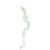
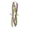
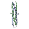
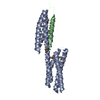
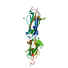
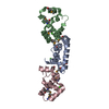
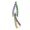
Journal: J Biol Chem / Year: 2016 Title: The Structure of the Plakin Domain of Plectin Reveals an Extended Rod-like Shape. Authors: Esther Ortega / José A Manso / Rubén M Buey / Ana M Carballido / Arturo Carabias / Arnoud Sonnenberg / José M de Pereda / Abstract: Plakins are large multi-domain proteins that interconnect cytoskeletal structures. Plectin is a prototypical plakin that tethers intermediate filaments to membrane-associated complexes. Most plakins ...Plakins are large multi-domain proteins that interconnect cytoskeletal structures. Plectin is a prototypical plakin that tethers intermediate filaments to membrane-associated complexes. Most plakins contain a plakin domain formed by up to nine spectrin repeats (SR1-SR9) and an SH3 domain. The plakin domains of plectin and other plakins harbor binding sites for junctional proteins. We have combined x-ray crystallography with small angle x-ray scattering (SAXS) to elucidate the structure of the plakin domain of plectin, extending our previous analysis of the SR1 to SR5 region. Two crystal structures of the SR5-SR6 region allowed us to characterize its uniquely wide inter-repeat conformational variability. We also report the crystal structures of the SR7-SR8 region, refined to 1.8 Å, and the SR7-SR9 at lower resolution. The SR7-SR9 region, which is conserved in all other plakin domains, forms a rigid segment stabilized by uniquely extensive inter-repeat contacts mediated by unusually long helices in SR8 and SR9. Using SAXS we show that in solution the SR3-SR6 and SR7-SR9 regions are rod-like segments and that SR3-SR9 of plectin has an extended shape with a small central kink. Other plakins, such as bullous pemphigoid antigen 1 and microtubule and actin cross-linking factor 1, are likely to have similar extended plakin domains. In contrast, desmoplakin has a two-segment structure with a central flexible hinge. The continuous versus segmented structures of the plakin domains of plectin and desmoplakin give insight into how different plakins might respond to tension and transmit mechanical signals.
Mass: 22323.803 Da / Num. of mol.: 2 / Mutation: F752A,F752A Source method: isolated from a genetically manipulated source Details: The SH3 domain, residues 819-888, has been replaced by the short sequence GSG Source: (gene. exp.) Homo sapiens (human) / Gene: PLEC, PLEC1 / Plasmid: MODIFIED pET15b / Production host: Escherichia coli BL21(DE3) (bacteria) / References: UniProt: Q15149
-
Experimental details
-
Experiment
Experiment
Method: X-RAY DIFFRACTION / Number of used crystals: 1
-
Sample preparation
Crystal
Density Matthews: 2.97 Å3/Da / Density % sol: 58 %
Crystal grow
Temperature: 277 K / Method: vapor diffusion, hanging drop / pH: 6 Details: A protein solution at 30 mg/ml in 10 mM Tris-Hcl (pH 7.5), 50 mM NaCl, 1 mM DTT was mixed with an equal volume of crystallization solution 0.1 M Bis-Tris-Propane (pH 6.0), 18% PEG 3350, 0.2 Na malonate
-
Data collection
Diffraction
Mean temperature: 100 K
Diffraction source
Source: ROTATING ANODE / Type: BRUKER AXS MICROSTAR-H / Wavelength: 1.5418 Å
Detector
Type: MAR scanner 345 mm plate / Detector: IMAGE PLATE / Date: Oct 8, 2010
Radiation
Monochromator: HELIOS OPTICS / Protocol: SINGLE WAVELENGTH / Monochromatic (M) / Laue (L): M / Scattering type: x-ray
Radiation wavelength
Wavelength: 1.5418 Å / Relative weight: 1
Reflection
Resolution: 3→43.4 Å / Num. obs: 10510 / % possible obs: 97.7 % / Observed criterion σ(I): -3 / Redundancy: 3.9 % / Biso Wilson estimate: 66.8 Å2 / CC1/2: 0.998 / Rpim(I) all: 0.093 / Net I/σ(I): 15
Reflection shell
Resolution: 3→3.08 Å / Redundancy: 3.9 % / Mean I/σ(I) obs: 2 / Rpim(I) all: 0.889 / % possible all: 100
-
Processing
Software
Name
Version
Classification
PHENIX
(dev_2000: ???)
refinement
XDS
January10, 2014
datareduction
XSCALE
January10, 2014
datascaling
PHASER
2.1.4
phasing
Refinement
Method to determine structure: MOLECULAR REPLACEMENT Starting model: D_1000218819 Resolution: 3→43.377 Å / SU ML: 0.48 / Cross valid method: FREE R-VALUE / σ(F): 1.36 / Phase error: 35.62
Rfactor
Num. reflection
% reflection
Selection details
Rfree
0.2772
510
4.86 %
0
Rwork
0.2447
-
-
-
obs
0.2464
10499
98.14 %
-
Solvent computation
Shrinkage radii: 0.8 Å / VDW probe radii: 1.1 Å
Displacement parameters
Biso mean: 73.3 Å2
Refinement step
Cycle: LAST / Resolution: 3→43.377 Å
Protein
Nucleic acid
Ligand
Solvent
Total
Num. atoms
2942
0
0
0
2942
Refine LS restraints
Refine-ID
Type
Dev ideal
Number
X-RAY DIFFRACTION
f_bond_d
0.002
2996
X-RAY DIFFRACTION
f_angle_d
0.563
4047
X-RAY DIFFRACTION
f_dihedral_angle_d
15.056
1083
X-RAY DIFFRACTION
f_chiral_restr
0.025
437
X-RAY DIFFRACTION
f_plane_restr
0.001
536
LS refinement shell
Resolution (Å)
Rfactor Rfree
Num. reflection Rfree
Rfactor Rwork
Num. reflection Rwork
Refine-ID
% reflection obs (%)
3.0002-3.302
0.4128
123
0.3628
2450
X-RAY DIFFRACTION
98
3.302-3.7796
0.2954
142
0.2589
2438
X-RAY DIFFRACTION
98
3.7796-4.761
0.2563
120
0.2292
2512
X-RAY DIFFRACTION
98
4.761-43.3818
0.2464
125
0.2146
2589
X-RAY DIFFRACTION
99
Refinement TLS params.
Method: refined / Refine-ID: X-RAY DIFFRACTION
ID
L11 (°2)
L12 (°2)
L13 (°2)
L22 (°2)
L23 (°2)
L33 (°2)
S11 (Å °)
S12 (Å °)
S13 (Å °)
S21 (Å °)
S22 (Å °)
S23 (Å °)
S31 (Å °)
S32 (Å °)
S33 (Å °)
T11 (Å2)
T12 (Å2)
T13 (Å2)
T22 (Å2)
T23 (Å2)
T33 (Å2)
Origin x (Å)
Origin y (Å)
Origin z (Å)
1
2.4057
-0.4734
2.6033
0.6199
-0.3665
2.6215
0.1949
-0.3628
-0.099
0.0483
-0.0106
0.1257
-0.026
-0.0958
0.0078
0.4
0.1095
-0.0141
0.3845
0.0514
0.475
30.6034
-17.923
6.1344
2
0.2819
-0.6084
0.7231
1.5216
-1.363
2.1644
0.4098
0.3975
0.6925
0.0326
-0.225
0.0394
0.313
-0.0289
0.0434
0.5004
0.1117
-0.0228
0.5701
0.0991
0.5736
14.5937
-11.2508
-4.8089
3
2.037
1.4248
0.3458
3.1092
-0.5679
1.5215
0.1553
-0.5552
0.5365
-0.2731
0.2054
0.4705
-0.422
-0.417
0.0035
0.4581
0.0827
-0.0391
0.729
0.0459
0.4895
0.4791
-3.3523
-13.254
4
1.5906
-2.0684
-1.6035
2.7421
2.5591
2.0155
0.0027
-0.2146
-0.1391
-0.2806
0.2124
-0.1538
-0.3814
0.4783
-0.0002
0.81
-0.0143
0.0871
0.657
-0.0969
0.5651
25.227
-21.3125
-49.51
5
2.9178
1.3791
-1.2168
2.1902
0.2476
4.5828
0.0455
0.3678
0.548
0.1875
-0.1321
0.0725
-0.0456
-0.3469
-0.0002
0.4444
-0.0114
-0.0446
0.3392
0.0292
0.3522
10.379
3.2134
-27.7297
Refinement TLS group
ID
Refine-ID
Refine TLS-ID
Selection details
1
X-RAY DIFFRACTION
1
chain 'A' and (resid746:821 )
2
X-RAY DIFFRACTION
2
chain 'A' and (resid889:945 )
3
X-RAY DIFFRACTION
3
chain 'A' and (resid946:1000 )
4
X-RAY DIFFRACTION
4
chain 'B' and (resid747:934 )
5
X-RAY DIFFRACTION
5
chain 'B' and (resid935:1001 )
+
About Yorodumi
-
News
-
Feb 9, 2022. New format data for meta-information of EMDB entries
New format data for meta-information of EMDB entries
Version 3 of the EMDB header file is now the official format.
The previous official version 1.9 will be removed from the archive.
In the structure databanks used in Yorodumi, some data are registered as the other names, "COVID-19 virus" and "2019-nCoV". Here are the details of the virus and the list of structure data.
Jan 31, 2019. EMDB accession codes are about to change! (news from PDBe EMDB page)
EMDB accession codes are about to change! (news from PDBe EMDB page)
The allocation of 4 digits for EMDB accession codes will soon come to an end. Whilst these codes will remain in use, new EMDB accession codes will include an additional digit and will expand incrementally as the available range of codes is exhausted. The current 4-digit format prefixed with “EMD-” (i.e. EMD-XXXX) will advance to a 5-digit format (i.e. EMD-XXXXX), and so on. It is currently estimated that the 4-digit codes will be depleted around Spring 2019, at which point the 5-digit format will come into force.
The EM Navigator/Yorodumi systems omit the EMD- prefix.
Related info.:Q: What is EMD? / ID/Accession-code notation in Yorodumi/EM Navigator
Yorodumi is a browser for structure data from EMDB, PDB, SASBDB, etc.
This page is also the successor to EM Navigator detail page, and also detail information page/front-end page for Omokage search.
The word "yorodu" (or yorozu) is an old Japanese word meaning "ten thousand". "mi" (miru) is to see.
Related info.:EMDB / PDB / SASBDB / Comparison of 3 databanks / Yorodumi Search / Aug 31, 2016. New EM Navigator & Yorodumi / Yorodumi Papers / Jmol/JSmol / Function and homology information / Changes in new EM Navigator and Yorodumi
 Movie
Movie Controller

 Yorodumi
 Open data
 Basic information
 Components
 Keywords
 Function and homology information
 Homo sapiens (human)
 X-RAY DIFFRACTION /
  Authors
 Spain, 1items
  Citation
 Structure visualization
 Downloads & links
 Other downloads


 PDBj
PDBj


 Assembly
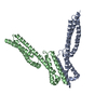


 Sample preparation
 Processing