[English] 日本語
Yorodumi
- PDB-5j1h: Structure of the spectrin repeats 5 and 6 of the plakin domain of... -

+
Open data


ID or keywords:

Loading...

-
Basic information

Entry
Database: PDB / ID: 5j1h
TitleStructure of the spectrin repeats 5 and 6 of the plakin domain of plectin
ComponentsPlectin,Plectin
KeywordsSTRUCTURAL PROTEIN / CYTOSKELETON / PLAKIN / INTERMEDIATE FILAMENT
Function / homology
Function and homology information


protein-containing complex organization / actomyosin contractile ring assembly actin filament organization / skeletal myofibril assembly / tight junction organization / Type I hemidesmosome assembly / hemidesmosome assembly / hemidesmosome / leukocyte migration involved in immune response / intermediate filament organization / : ...protein-containing complex organization / actomyosin contractile ring assembly actin filament organization / skeletal myofibril assembly / tight junction organization / Type I hemidesmosome assembly / hemidesmosome assembly / hemidesmosome / leukocyte migration involved in immune response / intermediate filament organization / : / intermediate filament cytoskeleton organization / regulation of vascular permeability / dystroglycan binding / cellular response to hydrostatic pressure / fibroblast migration / costamere / T cell chemotaxis / cellular response to fluid shear stress / peripheral nervous system myelin maintenance / cardiac muscle cell development / adherens junction organization / myoblast differentiation / intermediate filament cytoskeleton / response to food / structural constituent of muscle / ankyrin binding / intermediate filament / Assembly of collagen fibrils and other multimeric structures / sarcomere organization / nucleus organization / keratinocyte development / transmission of nerve impulse / brush border / sarcoplasm / Caspase-mediated cleavage of cytoskeletal proteins / establishment of skin barrier / skeletal muscle fiber development / respiratory electron transport chain / mitochondrion organization / wound healing / cell morphogenesis / multicellular organism growth / protein localization / structural constituent of cytoskeleton / sarcolemma / Z disc / cellular response to mechanical stimulus / : / actin filament binding / myelin sheath / gene expression / mitochondrial outer membrane / cadherin binding / axon / focal adhesion / dendrite / perinuclear region of cytoplasm / RNA binding / extracellular exosome / plasma membrane / cytoplasm / cytosol
Similarity search - Function
: / Spectrin-like repeat / Spectrin repeat / Spectrin-like repeat / Desmoplakin, spectrin-like domain / Spectrin like domain / Plectin repeat / Plectin repeat / Plakin repeat superfamily / Desmoplakin, SH3 domain ...: / Spectrin-like repeat / Spectrin repeat / Spectrin-like repeat / Desmoplakin, spectrin-like domain / Spectrin like domain / Plectin repeat / Plectin repeat / Plakin repeat superfamily / Desmoplakin, SH3 domain / SH3 domain / Plectin repeat / Plakin / Spectrin/alpha-actinin / Actinin-type actin-binding domain signature 1. / Actinin-type actin-binding domain signature 2. / Spectrin repeats / Actinin-type actin-binding domain, conserved site / Calponin homology domain / Calponin homology (CH) domain / Calponin homology domain / CH domain superfamily / Calponin homology (CH) domain profile. / Plectin/S10, N-terminal / Plectin/S10 domain / Src homology 3 (SH3) domain profile. / SH3 domain / Winged helix-like DNA-binding domain superfamily
Similarity search - Domain/homology
Biological speciesHomo sapiens (human)
MethodX-RAY DIFFRACTION / SYNCHROTRON / SIRAS / Resolution: 2.8 Å
AuthorsOrtega, E. / DE PEREDA, J.M.
Funding support Spain, 1items
OrganizationGrant numberCountry
Spanish Ministry of Economy and CompetitivenessBFU2012-32847 Spain
Citation
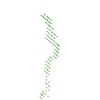
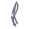
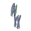
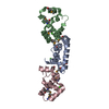
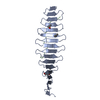
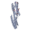
Journal: J Biol Chem / Year: 2016
Title: The Structure of the Plakin Domain of Plectin Reveals an Extended Rod-like Shape.
Authors: Esther Ortega / José A Manso / Rubén M Buey / Ana M Carballido / Arturo Carabias / Arnoud Sonnenberg / José M de Pereda /
Abstract: Plakins are large multi-domain proteins that interconnect cytoskeletal structures. Plectin is a prototypical plakin that tethers intermediate filaments to membrane-associated complexes. Most plakins ...Plakins are large multi-domain proteins that interconnect cytoskeletal structures. Plectin is a prototypical plakin that tethers intermediate filaments to membrane-associated complexes. Most plakins contain a plakin domain formed by up to nine spectrin repeats (SR1-SR9) and an SH3 domain. The plakin domains of plectin and other plakins harbor binding sites for junctional proteins. We have combined x-ray crystallography with small angle x-ray scattering (SAXS) to elucidate the structure of the plakin domain of plectin, extending our previous analysis of the SR1 to SR5 region. Two crystal structures of the SR5-SR6 region allowed us to characterize its uniquely wide inter-repeat conformational variability. We also report the crystal structures of the SR7-SR8 region, refined to 1.8 Å, and the SR7-SR9 at lower resolution. The SR7-SR9 region, which is conserved in all other plakin domains, forms a rigid segment stabilized by uniquely extensive inter-repeat contacts mediated by unusually long helices in SR8 and SR9. Using SAXS we show that in solution the SR3-SR6 and SR7-SR9 regions are rod-like segments and that SR3-SR9 of plectin has an extended shape with a small central kink. Other plakins, such as bullous pemphigoid antigen 1 and microtubule and actin cross-linking factor 1, are likely to have similar extended plakin domains. In contrast, desmoplakin has a two-segment structure with a central flexible hinge. The continuous versus segmented structures of the plakin domains of plectin and desmoplakin give insight into how different plakins might respond to tension and transmit mechanical signals.
History
DepositionMar 29, 2016Deposition site: RCSB / Processing site: PDBE
Revision 1.0Jul 20, 2016Provider: repository / Type: Initial release
Revision 1.1Jul 27, 2016Group: Database references
Revision 1.2Sep 14, 2016Group: Database references
Revision 1.3Sep 6, 2017Group: Author supporting evidence / Category: pdbx_audit_support / Item: _pdbx_audit_support.funding_organization
Revision 1.4Jun 12, 2019Group: Data collection / Structure summary
Category: audit_author / database_PDB_rev / database_PDB_rev_record
Item: _audit_author.name
Revision 1.5May 8, 2024Group: Data collection / Database references / Category: chem_comp_atom / chem_comp_bond / database_2
Item: _database_2.pdbx_DOI / _database_2.pdbx_database_accession

-
Structure visualization

Structure viewerMolecule:
MolmilJmol/JSmol

Downloads & links

-
Assembly

Deposited unit
A: Plectin,Plectin
B: Plectin,Plectin


Theoretical massNumber of molelcules
Total (without water)44,9362
Polymers44,9362
Non-polymers00
Water362
1
A: Plectin,Plectin


Theoretical massNumber of molelcules
Total (without water)22,4681
Polymers22,4681
Non-polymers00
Water181
TypeNameSymmetry operationNumber
identity operation1_555x,y,z1
2
B: Plectin,Plectin


Theoretical massNumber of molelcules
Total (without water)22,4681
Polymers22,4681
Non-polymers00
Water0
TypeNameSymmetry operationNumber
identity operation1_555x,y,z1
Unit cell
Length a, b, c (Å)178.140, 178.140, 50.680
Angle α, β, γ (deg.)90.00, 90.00, 120.00
Int Tables number146
Space group name H-MH3

-
Components

#1: Protein Plectin,Plectin / PLTN / Hemidesmosomal protein 1 / HD1 / Plectin-1


Mass: 22467.932 Da / Num. of mol.: 2 / Mutation: F752A,F752A
Source method: isolated from a genetically manipulated source
Details: The residues 819-888 that code for an SH3 domain have been replaced by the Gly-Ser sequence GSGSG
Source: (gene. exp.) Homo sapiens (human) / Gene: PLEC, PLEC1 / Plasmid: MODIFIED pET15b / Production host: Escherichia coli BL21(DE3) (bacteria) / References: UniProt: Q15149
#2: Water ChemComp-HOH / water


Mass: 18.015 Da / Num. of mol.: 2 / Source method: isolated from a natural source / Formula: H2O

-
Experimental details

-
Experiment

ExperimentMethod: X-RAY DIFFRACTION / Number of used crystals: 1

-
Sample preparation

CrystalDensity Matthews: 3.33 Å3/Da / Density % sol: 63 %
Crystal growTemperature: 277 K / Method: vapor diffusion, sitting drop / pH: 7.5
Details: Protein at 30 mg/ml in 10 mM Tris-HCl (pH 7.5), 50 mM NaCl, 1 mM DTT was mixed with an equal volume of the crystallization solution 0.2 M Na/K tartrate, 20% PEG3350

-
Data collection

DiffractionMean temperature: 105 K
Diffraction sourceSource: SYNCHROTRON / Site: SLS / Beamline: X06DA / Wavelength: 1 Å
DetectorType: DECTRIS PILATUS 2M-F / Detector: PIXEL / Date: Nov 14, 2012
RadiationProtocol: SINGLE WAVELENGTH / Monochromatic (M) / Laue (L): M / Scattering type: x-ray
Radiation wavelengthWavelength: 1 Å / Relative weight: 1
ReflectionResolution: 2.8→48.149 Å / Num. obs: 14753 / % possible obs: 100 % / Observed criterion σ(I): -3 / Redundancy: 10.2 % / Biso Wilson estimate: 55.6 Å2 / CC1/2: 0.999 / Rpim(I) all: 0.122 / Net I/σ(I): 27.8
Reflection shellResolution: 2.8→2.87 Å / Redundancy: 10.7 % / Mean I/σ(I) obs: 2 / Rpim(I) all: 2.21 / % possible all: 100

-
Processing

Software
NameVersionClassification
PHENIXdev_1931refinement
XDSNovember 3, 2014data reduction
XSCALENovember 3, 2014data scaling
SHELXDEVersion 2013/3phasing
autoSHARPphasing
RefinementMethod to determine structure: SIRAS / Resolution: 2.8→48.149 Å / SU ML: 0.28 / Cross valid method: FREE R-VALUE / σ(F): 1.94 / Phase error: 29.95
RfactorNum. reflection% reflectionSelection details
Rfree0.2693 714 4.86 %0
Rwork0.2226 ---
obs0.2247 14690 99.55 %-
Solvent computationShrinkage radii: 0.9 Å / VDW probe radii: 1.11 Å
Displacement parametersBiso mean: 90.1 Å2
Refinement stepCycle: LAST / Resolution: 2.8→48.149 Å
ProteinNucleic acidLigandSolventTotal
Num. atoms2657 0 0 2 2659
Refine LS restraints
Refine-IDTypeDev idealNumber
X-RAY DIFFRACTIONf_bond_d0.0022705
X-RAY DIFFRACTIONf_angle_d0.4483663
X-RAY DIFFRACTIONf_dihedral_angle_d13.001928
X-RAY DIFFRACTIONf_chiral_restr0.022414
X-RAY DIFFRACTIONf_plane_restr0.001484
LS refinement shell
Resolution (Å)Rfactor RfreeNum. reflection RfreeRfactor RworkNum. reflection RworkRefine-ID% reflection obs (%)
2.8002-3.01640.3211520.29932768X-RAY DIFFRACTION99
3.0164-3.31980.2651390.25292769X-RAY DIFFRACTION99
3.3198-3.80010.26151320.21712834X-RAY DIFFRACTION100
3.8001-4.7870.24411500.20652791X-RAY DIFFRACTION100
4.787-48.15560.28471410.21122814X-RAY DIFFRACTION100
Refinement TLS params.

Method: refined / Refine-ID: X-RAY DIFFRACTION

IDL112)L122)L132)L222)L232)L332)S11 (Å °)S12 (Å °)S13 (Å °)S21 (Å °)S22 (Å °)S23 (Å °)S31 (Å °)S32 (Å °)S33 (Å °)T112)T122)T132)T222)T232)T332)Origin x (Å)Origin y (Å)Origin z (Å)
11.1214-0.5641-0.4571.38020.30370.19410.10310.3219-0.93850.10380.0987-1.1046-0.0697-0.11480.00670.6731-0.0966-0.17580.6526-0.0730.949443.271714.840921.7459
21.8906-0.78440.0851.74950.49470.94420.07350.2311-0.77540.29710.0857-0.7616-0.0734-0.06340.01710.4184-0.0601-0.05790.555-0.00320.473936.884524.905926.2539
34.9018-0.3310.12421.61871.15753.3256-0.03630.20030.25290.1488-0.1828-0.4132-0.462-0.0253-0.00850.6592-0.0193-0.04710.48080.06130.105532.788652.753927.3188
40.9451-0.1547-0.05770.46390.10830.3038-0.0080.2144-0.4516-0.0997-0.1415-0.48380.15980.11330.09680.1769-0.06390.10130.5451-0.10861.896867.32253.019511.5949
50.8041-0.5155-0.12090.41270.36671.60850.0746-0.06130.3738-0.21350.2073-1.4679-0.64830.3107-0.08970.4045-0.10240.21930.7618-0.03881.742866.585120.136514.6153
60.3249-0.22070.18190.1544-0.11920.11980.0464-0.02811.2314-0.94820.3478-1.832-0.8193-0.244-0.02371.3155-0.17260.52261.2439-0.07722.270260.922548.52416.304
Refinement TLS group
IDRefine-IDRefine TLS-IDSelection details
1X-RAY DIFFRACTION1chain 'A' and (resid 750 through 887 )
2X-RAY DIFFRACTION2chain 'A' and (resid 888 through 934 )
3X-RAY DIFFRACTION3chain 'A' and (resid 935 through 1000 )
4X-RAY DIFFRACTION4chain 'B' and (resid 750 through 775 )
5X-RAY DIFFRACTION5chain 'B' and (resid 776 through 934 )
6X-RAY DIFFRACTION6chain 'B' and (resid 935 through 996 )

+
About Yorodumi

-
News

-
Feb 9, 2022. New format data for meta-information of EMDB entries

New format data for meta-information of EMDB entries

  • Version 3 of the EMDB header file is now the official format.
  • The previous official version 1.9 will be removed from the archive.

Related info.:EMDB header

External links:wwPDB to switch to version 3 of the EMDB data model

-
Aug 12, 2020. Covid-19 info

Covid-19 info

URL: https://pdbjlvh1.pdbj.org/emnavi/covid19.php

New page: Covid-19 featured information page in EM Navigator.

Related info.:Covid-19 info / Mar 5, 2020. Novel coronavirus structure data

+
Mar 5, 2020. Novel coronavirus structure data

Novel coronavirus structure data

Related info.:Yorodumi Speices / Aug 12, 2020. Covid-19 info

External links:COVID-19 featured content - PDBj / Molecule of the Month (242):Coronavirus Proteases

+
Jan 31, 2019. EMDB accession codes are about to change! (news from PDBe EMDB page)

EMDB accession codes are about to change! (news from PDBe EMDB page)

  • The allocation of 4 digits for EMDB accession codes will soon come to an end. Whilst these codes will remain in use, new EMDB accession codes will include an additional digit and will expand incrementally as the available range of codes is exhausted. The current 4-digit format prefixed with “EMD-” (i.e. EMD-XXXX) will advance to a 5-digit format (i.e. EMD-XXXXX), and so on. It is currently estimated that the 4-digit codes will be depleted around Spring 2019, at which point the 5-digit format will come into force.
  • The EM Navigator/Yorodumi systems omit the EMD- prefix.

Related info.:Q: What is EMD? / ID/Accession-code notation in Yorodumi/EM Navigator

External links:EMDB Accession Codes are Changing Soon! / Contact to PDBj

+
Jul 12, 2017. Major update of PDB

Major update of PDB

  • wwPDB released updated PDB data conforming to the new PDBx/mmCIF dictionary.
  • This is a major update changing the version number from 4 to 5, and with Remediation, in which all the entries are updated.
  • In this update, many items about electron microscopy experimental information are reorganized (e.g. em_software).
  • Now, EM Navigator and Yorodumi are based on the updated data.

External links:wwPDB Remediation / Enriched Model Files Conforming to OneDep Data Standards Now Available in the PDB FTP Archive

-
Yorodumi

Thousand views of thousand structures

  • Yorodumi is a browser for structure data from EMDB, PDB, SASBDB, etc.
  • This page is also the successor to EM Navigator detail page, and also detail information page/front-end page for Omokage search.
  • The word "yorodu" (or yorozu) is an old Japanese word meaning "ten thousand". "mi" (miru) is to see.

Related info.:EMDB / PDB / SASBDB / Comparison of 3 databanks / Yorodumi Search / Aug 31, 2016. New EM Navigator & Yorodumi / Yorodumi Papers / Jmol/JSmol / Function and homology information / Changes in new EM Navigator and Yorodumi

Read more