 Movie
Movie Controller

[English] 日本語
 Yorodumi
- PDB-5j1i: Structure of the spectrin repeats 7, 8, and 9 of the plakin domai... -
+ Open data

- Basic information
| Entry | Database: PDB / ID: 5j1i | ||||||
|---|---|---|---|---|---|---|---|
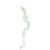
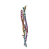
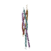
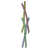
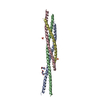
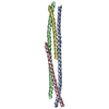
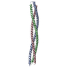
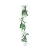
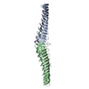
| Title | Structure of the spectrin repeats 7, 8, and 9 of the plakin domain of plectin | ||||||
 Components Components | Plectin | ||||||
 Keywords Keywords | STRUCTURAL PROTEIN / CYTOSKELETON / PLAKIN / INTERMEDIATE FILAMENT | ||||||
| Function / homology |  Function and homology information Function and homology informationactomyosin contractile ring assembly actin filament organization / protein-containing complex organization / tight junction organization / Type I hemidesmosome assembly / skeletal myofibril assembly / hemidesmosome assembly / hemidesmosome / leukocyte migration involved in immune response / intermediate filament organization / fibroblast migration ...actomyosin contractile ring assembly actin filament organization / protein-containing complex organization / tight junction organization / Type I hemidesmosome assembly / skeletal myofibril assembly / hemidesmosome assembly / hemidesmosome / leukocyte migration involved in immune response / intermediate filament organization / fibroblast migration / intermediate filament cytoskeleton organization / dystroglycan binding / cellular response to fluid shear stress / cellular response to hydrostatic pressure / costamere / T cell chemotaxis / adherens junction organization / peripheral nervous system myelin maintenance / intermediate filament cytoskeleton / myoblast differentiation / regulation of vascular permeability / podosome / cardiac muscle cell development / sarcomere organization / Assembly of collagen fibrils and other multimeric structures / nucleus organization / ankyrin binding / structural constituent of muscle / keratinocyte development / response to food / establishment of skin barrier / transmission of nerve impulse / sarcoplasm / brush border / Caspase-mediated cleavage of cytoskeletal proteins / skeletal muscle fiber development / multicellular organism growth / respiratory electron transport chain / mitochondrion organization / wound healing / cellular response to mechanical stimulus / sarcolemma / structural constituent of cytoskeleton / cell morphogenesis / Z disc / actin filament binding / intracellular protein localization / myelin sheath / gene expression / mitochondrial outer membrane / cadherin binding / axon / focal adhesion / dendrite / perinuclear region of cytoplasm / RNA binding / extracellular exosome / identical protein binding / plasma membrane / cytosol / cytoplasm Similarity search - Function | ||||||
| Biological species |  Homo sapiens (human) Homo sapiens (human) | ||||||
| Method |  X-RAY DIFFRACTION / SYNCHROTRON / MOLECULAR REPLACEMENT / Resolution: 2.801 Å X-RAY DIFFRACTION / SYNCHROTRON / MOLECULAR REPLACEMENT / Resolution: 2.801 Å | ||||||
 Authors Authors | Ortega, E. / DE PEREDA, J.M. | ||||||
| Funding support |  Spain, 1items Spain, 1items
| ||||||
 Citation Citation | Journal: J Biol Chem / Year: 2016 Title: The Structure of the Plakin Domain of Plectin Reveals an Extended Rod-like Shape. Authors: Esther Ortega / José A Manso / Rubén M Buey / Ana M Carballido / Arturo Carabias / Arnoud Sonnenberg / José M de Pereda /  Abstract: Plakins are large multi-domain proteins that interconnect cytoskeletal structures. Plectin is a prototypical plakin that tethers intermediate filaments to membrane-associated complexes. Most plakins ...Plakins are large multi-domain proteins that interconnect cytoskeletal structures. Plectin is a prototypical plakin that tethers intermediate filaments to membrane-associated complexes. Most plakins contain a plakin domain formed by up to nine spectrin repeats (SR1-SR9) and an SH3 domain. The plakin domains of plectin and other plakins harbor binding sites for junctional proteins. We have combined x-ray crystallography with small angle x-ray scattering (SAXS) to elucidate the structure of the plakin domain of plectin, extending our previous analysis of the SR1 to SR5 region. Two crystal structures of the SR5-SR6 region allowed us to characterize its uniquely wide inter-repeat conformational variability. We also report the crystal structures of the SR7-SR8 region, refined to 1.8 Å, and the SR7-SR9 at lower resolution. The SR7-SR9 region, which is conserved in all other plakin domains, forms a rigid segment stabilized by uniquely extensive inter-repeat contacts mediated by unusually long helices in SR8 and SR9. Using SAXS we show that in solution the SR3-SR6 and SR7-SR9 regions are rod-like segments and that SR3-SR9 of plectin has an extended shape with a small central kink. Other plakins, such as bullous pemphigoid antigen 1 and microtubule and actin cross-linking factor 1, are likely to have similar extended plakin domains. In contrast, desmoplakin has a two-segment structure with a central flexible hinge. The continuous versus segmented structures of the plakin domains of plectin and desmoplakin give insight into how different plakins might respond to tension and transmit mechanical signals. | ||||||
| History |
|
- Structure visualization
| Structure viewer | Molecule: MolmilJmol/JSmol |
|---|
- Downloads & links
-Download
| PDBx/mmCIF format | 5j1i.cif.gz | 264.5 KB | Display | PDBx/mmCIF format |
|---|---|---|---|---|
| PDB format | pdb5j1i.ent.gz | 215.1 KB | Display | PDB format |
| PDBx/mmJSON format | 5j1i.json.gz | Tree view | PDBx/mmJSON format | |
| Others |  Other downloads Other downloads |
-Validation report
| Arichive directory | https://data.pdbj.org/pub/pdb/validation_reports/j1/5j1iftp://data.pdbj.org/pub/pdb/validation_reports/j1/5j1i | HTTPS FTP |
|---|
-Related structure data
| Related structure data |  5j1fC  5j1gSC  5j1hC S: Starting model for refinement C: citing same article ( |
|---|---|
| Similar structure data | |
| Experimental dataset #1 | Data reference: 10.5281/zenodo.814209 / Data set type: diffraction image data |


-Links
 PDBj
PDBj


- Assembly
| Deposited unit | 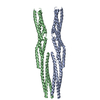
| ||||||||
|---|---|---|---|---|---|---|---|---|---|
| 1 | 
| ||||||||
| 2 | 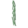
| ||||||||
| Unit cell |
|
-Components
| #1: Protein | Mass: 42889.562 Da / Num. of mol.: 2 / Fragment: UNP residues 1104-1372 Source method: isolated from a genetically manipulated source Source: (gene. exp.) Homo sapiens (human) / Gene: PLEC, PLEC1 / Plasmid: MODIFIED pET15b / Production host:  |
|---|
-Experimental details
-Experiment
| Experiment | Method: X-RAY DIFFRACTION / Number of used crystals: 1 |
|---|
- Sample preparation
| Crystal | Density Matthews: 3.04 Å3/Da / Density % sol: 60 % |
|---|---|
| Crystal grow | Temperature: 298 K / Method: vapor diffusion, hanging drop / pH: 8 Details: Protein solution at 12 mg/ml in 10 mM Tris-HCl (pH 7.5), 50 mM NaCl, 1 mM DTT was mixed with crystallization solution 0.1 M Bis-Tris-propane (pH 8.0), 16% PEG 3350, 0.3 M Na/K tartrate |
-Data collection
| Diffraction | Mean temperature: 100 K |
|---|---|
| Diffraction source | Source: SYNCHROTRON / Site: ESRF  / Beamline: ID14-2 / Wavelength: 0.933 Å / Beamline: ID14-2 / Wavelength: 0.933 Å |
| Detector | Type: ADSC QUANTUM 4 / Detector: CCD / Date: Dec 10, 2009 |
| Radiation | Protocol: SINGLE WAVELENGTH / Monochromatic (M) / Laue (L): M / Scattering type: x-ray |
| Radiation wavelength | Wavelength: 0.933 Å / Relative weight: 1 |
| Reflection | Resolution: 2.8→48.878 Å / Num. obs: 14129 / % possible obs: 55.7 % / Observed criterion σ(I): -3 / Redundancy: 7.5 % / CC1/2: 1 / Rpim(I) all: 0.083 / Net I/σ(I): 17.2 |
| Reflection shell | Resolution: 2.8→2.87 Å / Redundancy: 6.2 % / Mean I/σ(I) obs: 1.7 / Rpim(I) all: 1.38 / % possible all: 11.4 |
- Processing
| Software |
| |||||||||||||||||||||||||||||||||||||||||||||||||||||||||||||||||||||||||||
|---|---|---|---|---|---|---|---|---|---|---|---|---|---|---|---|---|---|---|---|---|---|---|---|---|---|---|---|---|---|---|---|---|---|---|---|---|---|---|---|---|---|---|---|---|---|---|---|---|---|---|---|---|---|---|---|---|---|---|---|---|---|---|---|---|---|---|---|---|---|---|---|---|---|---|---|---|
| Refinement | Method to determine structure: MOLECULAR REPLACEMENT Starting model: 5J1G Resolution: 2.801→48.878 Å / SU ML: 0.46 / Cross valid method: FREE R-VALUE / σ(F): 1.36 / Phase error: 39.29 Details: Diffraction was highly anisotropic; the approximate resolution limits and the 3 main directions were: 2.8 A (-0.38 a* + 0.93 c*), 3.8 A (b*), and 5.0 A (0.53 a* + 0.85 c*). Refinement was ...Details: Diffraction was highly anisotropic; the approximate resolution limits and the 3 main directions were: 2.8 A (-0.38 a* + 0.93 c*), 3.8 A (b*), and 5.0 A (0.53 a* + 0.85 c*). Refinement was done against data corrected with the STARANISO server.
| |||||||||||||||||||||||||||||||||||||||||||||||||||||||||||||||||||||||||||
| Solvent computation | Shrinkage radii: 0.9 Å / VDW probe radii: 1.11 Å | |||||||||||||||||||||||||||||||||||||||||||||||||||||||||||||||||||||||||||
| Displacement parameters | Biso mean: 81.2 Å2 | |||||||||||||||||||||||||||||||||||||||||||||||||||||||||||||||||||||||||||
| Refinement step | Cycle: LAST / Resolution: 2.801→48.878 Å
| |||||||||||||||||||||||||||||||||||||||||||||||||||||||||||||||||||||||||||
| Refine LS restraints |
| |||||||||||||||||||||||||||||||||||||||||||||||||||||||||||||||||||||||||||
| LS refinement shell |
| |||||||||||||||||||||||||||||||||||||||||||||||||||||||||||||||||||||||||||
| Refinement TLS params. | Method: refined / Refine-ID: X-RAY DIFFRACTION
| |||||||||||||||||||||||||||||||||||||||||||||||||||||||||||||||||||||||||||
| Refinement TLS group |
|
