 Movie
Movie Controller

[English] 日本語
 Yorodumi
- PDB-4p9t: Structure of the free form of the N-terminal VH1 domain of monome... -
+ Open data

- Basic information
| Entry | Database: PDB / ID: 4p9t | ||||||
|---|---|---|---|---|---|---|---|
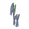
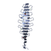
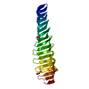
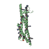
| Title | Structure of the free form of the N-terminal VH1 domain of monomeric alpha-catenin | ||||||
 Components Components | Catenin alpha-2 | ||||||
 Keywords Keywords | CELL ADHESION / Cytoskeletal protein / adherens junction / helix bundle | ||||||
| Function / homology |  Function and homology information Function and homology informationradial glia guided migration of Purkinje cell / regulation of synapse structural plasticity / extrinsic component of postsynaptic membrane / extrinsic component of presynaptic membrane / modification of postsynaptic actin cytoskeleton / presynaptic active zone cytoplasmic component / negative regulation of Arp2/3 complex-mediated actin nucleation / Myogenesis / catenin complex / brain morphogenesis ...radial glia guided migration of Purkinje cell / regulation of synapse structural plasticity / extrinsic component of postsynaptic membrane / extrinsic component of presynaptic membrane / modification of postsynaptic actin cytoskeleton / presynaptic active zone cytoplasmic component / negative regulation of Arp2/3 complex-mediated actin nucleation / Myogenesis / catenin complex / brain morphogenesis / regulation of neuron migration / dendrite morphogenesis / regulation of neuron projection development / parallel fiber to Purkinje cell synapse / prepulse inhibition / postsynaptic density, intracellular component / axonogenesis / hippocampal mossy fiber to CA3 synapse / adherens junction / cell-cell adhesion / beta-catenin binding / actin filament binding / cell migration / lamellipodium / actin cytoskeleton / basolateral plasma membrane / postsynaptic density / cadherin binding / axon / structural molecule activity / identical protein binding / nucleus / cytoplasm Similarity search - Function | ||||||
| Biological species |  | ||||||
| Method |  X-RAY DIFFRACTION / SYNCHROTRON / MOLECULAR REPLACEMENT / Resolution: 2.5 Å X-RAY DIFFRACTION / SYNCHROTRON / MOLECULAR REPLACEMENT / Resolution: 2.5 Å | ||||||
 Authors Authors | Shibahara, T. / Hirano, Y. / Hakoshima, T. | ||||||
 Citation Citation | Journal: Febs Lett. / Year: 2015 Title: Structure of the free form of the N-terminal VH1 domain of monomeric alpha-catenin. Authors: Shibahara, T. / Hirano, Y. / Hakoshima, T. | ||||||
| History |
|
- Structure visualization
| Structure viewer | Molecule: MolmilJmol/JSmol |
|---|
- Downloads & links
-Download
| PDBx/mmCIF format | 4p9t.cif.gz | 182.1 KB | Display | PDBx/mmCIF format |
|---|---|---|---|---|
| PDB format | pdb4p9t.ent.gz | 140.2 KB | Display | PDB format |
| PDBx/mmJSON format | 4p9t.json.gz | Tree view | PDBx/mmJSON format | |
| Others |  Other downloads Other downloads |
-Validation report
| Arichive directory | https://data.pdbj.org/pub/pdb/validation_reports/p9/4p9tftp://data.pdbj.org/pub/pdb/validation_reports/p9/4p9t | HTTPS FTP |
|---|
-Related structure data
| Related structure data | 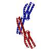 1dovS S: Starting model for refinement |
|---|---|
| Similar structure data |


-Links
 PDBj
PDBj

- Assembly
| Deposited unit | 
| |||||||||||||||||||||||||||||||||||||||||||||||||||||||||||||||||||||||||||||||||||||||||||
|---|---|---|---|---|---|---|---|---|---|---|---|---|---|---|---|---|---|---|---|---|---|---|---|---|---|---|---|---|---|---|---|---|---|---|---|---|---|---|---|---|---|---|---|---|---|---|---|---|---|---|---|---|---|---|---|---|---|---|---|---|---|---|---|---|---|---|---|---|---|---|---|---|---|---|---|---|---|---|---|---|---|---|---|---|---|---|---|---|---|---|---|---|
| 1 | 
| |||||||||||||||||||||||||||||||||||||||||||||||||||||||||||||||||||||||||||||||||||||||||||
| 2 | 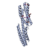
| |||||||||||||||||||||||||||||||||||||||||||||||||||||||||||||||||||||||||||||||||||||||||||
| 3 | 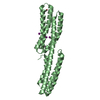
| |||||||||||||||||||||||||||||||||||||||||||||||||||||||||||||||||||||||||||||||||||||||||||
| 4 | 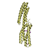
| |||||||||||||||||||||||||||||||||||||||||||||||||||||||||||||||||||||||||||||||||||||||||||
| Unit cell |
| |||||||||||||||||||||||||||||||||||||||||||||||||||||||||||||||||||||||||||||||||||||||||||
| Noncrystallographic symmetry (NCS) | NCS domain:
NCS domain segments: Ens-ID: 1
|