 Movie
Movie Controller


+ Open data

- Basic information
| Entry | Database: PDB / ID: 1dov | ||||||
|---|---|---|---|---|---|---|---|
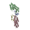
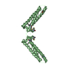
| Title | CRYSTAL STRUCTURE OF THE ALPHA-CATENIN DIMERIZATION DOMAIN | ||||||
 Components Components | ALPHA-CATENIN | ||||||
 Keywords Keywords | CELL ADHESION / four-helix bundle | ||||||
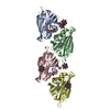
| Function / homology |  Function and homology information Function and homology informationnegative regulation of integrin-mediated signaling pathway / VEGFR2 mediated vascular permeability / Adherens junctions interactions / RHO GTPases activate IQGAPs / epithelial cell-cell adhesion / Regulation of CDH1 Function / zonula adherens / Degradation of CDH1 / gamma-catenin binding / gap junction assembly ...negative regulation of integrin-mediated signaling pathway / VEGFR2 mediated vascular permeability / Adherens junctions interactions / RHO GTPases activate IQGAPs / epithelial cell-cell adhesion / Regulation of CDH1 Function / zonula adherens / Degradation of CDH1 / gamma-catenin binding / gap junction assembly / cellular response to indole-3-methanol / vinculin binding / Myogenesis / flotillin complex / apical junction assembly / catenin complex / positive regulation of smoothened signaling pathway / positive regulation of extrinsic apoptotic signaling pathway in absence of ligand / negative regulation of cell motility / axon regeneration / negative regulation of protein localization to nucleus / negative regulation of neuroblast proliferation / odontogenesis of dentin-containing tooth / establishment or maintenance of cell polarity / intercalated disc / negative regulation of extrinsic apoptotic signaling pathway in absence of ligand / ovarian follicle development / acrosomal vesicle / adherens junction / cell-cell adhesion / male gonad development / response to estrogen / beta-catenin binding / cell junction / cell-cell junction / actin filament binding / intracellular protein localization / regulation of cell population proliferation / cell migration / lamellipodium / actin cytoskeleton / cadherin binding / negative regulation of apoptotic process / structural molecule activity / Golgi apparatus / identical protein binding / nucleus / plasma membrane Similarity search - Function | ||||||
| Biological species |  | ||||||
| Method |  X-RAY DIFFRACTION / SYNCHROTRON / Resolution: 3 Å X-RAY DIFFRACTION / SYNCHROTRON / Resolution: 3 Å | ||||||
 Authors Authors | Pokutta, S. / Weis, W.I. | ||||||
 Citation Citation | Journal: Mol.Cell / Year: 2000 Title: Structure of the dimerization and beta-catenin-binding region of alpha-catenin. Authors: Pokutta, S. / Weis, W.I. | ||||||
| History |
|
- Structure visualization
| Structure viewer | Molecule: MolmilJmol/JSmol |
|---|
- Downloads & links
-Download
| PDBx/mmCIF format | 1dov.cif.gz | 41.4 KB | Display | PDBx/mmCIF format |
|---|---|---|---|---|
| PDB format | pdb1dov.ent.gz | 30.6 KB | Display | PDB format |
| PDBx/mmJSON format | 1dov.json.gz | Tree view | PDBx/mmJSON format | |
| Others |  Other downloads Other downloads |
-Validation report
| Arichive directory | https://data.pdbj.org/pub/pdb/validation_reports/do/1dovftp://data.pdbj.org/pub/pdb/validation_reports/do/1dov | HTTPS FTP |
|---|
-Related structure data


-Links
 PDBj
PDBj

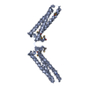
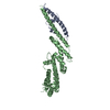
- Assembly
| Deposited unit | 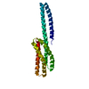
| ||||||||
|---|---|---|---|---|---|---|---|---|---|
| 1 | 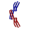
| ||||||||
| 2 | 
| ||||||||
| Unit cell |
| ||||||||
| Details | The biological assembly is a dimer. The twofold axis of the dimer is coincident with a crystallographic twofold. The dimer can by generated by application of the following rotation matrix: (-0.5 0.866 0.0) (0.866 0.5 0.0) (0.0 0.0 -1.0) and the translation vector (0. 0. 118.17) |
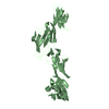
-Components
| #1: Protein | Mass: 19875.953 Da / Num. of mol.: 1 / Fragment: DIMERIZATION DOMAIN Source method: isolated from a genetically manipulated source Source: (gene. exp.)  |
|---|
-Experimental details
-Experiment
| Experiment | Method: X-RAY DIFFRACTION / Number of used crystals: 1 |
|---|
- Sample preparation
| Crystal | Density Matthews: 5.33 Å3/Da / Density % sol: 76.91 % | ||||||||||||||||||||||||||||||
|---|---|---|---|---|---|---|---|---|---|---|---|---|---|---|---|---|---|---|---|---|---|---|---|---|---|---|---|---|---|---|---|
| Crystal grow | Temperature: 295 K / Method: vapor diffusion, hanging drop / pH: 8.9 Details: PEG 400, Tris, urea, dithiothreitol, pH 8.9, VAPOR DIFFUSION, HANGING DROP, temperature 295K | ||||||||||||||||||||||||||||||
| Crystal grow | *PLUS | ||||||||||||||||||||||||||||||
| Components of the solutions | *PLUS
|
-Data collection
| Diffraction | Mean temperature: 288 K |
|---|---|
| Diffraction source | Source: SYNCHROTRON / Site: SSRL  / Beamline: BL9-1 / Wavelength: 0.98 / Beamline: BL9-1 / Wavelength: 0.98 |
| Detector | Type: MAR scanner 345 mm plate / Detector: IMAGE PLATE / Date: Nov 25, 1997 |
| Radiation | Protocol: SINGLE WAVELENGTH / Monochromatic (M) / Laue (L): M / Scattering type: x-ray |
| Radiation wavelength | Wavelength: 0.98 Å / Relative weight: 1 |
| Reflection | Resolution: 3→40 Å / Num. all: 33971 / Num. obs: 8574 / % possible obs: 99.6 % / Observed criterion σ(F): 0 / Observed criterion σ(I): 3 / Redundancy: 4 % / Rmerge(I) obs: 0.082 / Net I/σ(I): 11.6 |
| Reflection shell | Resolution: 3→3.16 Å / Redundancy: 4 % / Rmerge(I) obs: 0.322 / Num. unique all: 5031 / % possible all: 100 |
| Reflection | *PLUS Num. measured all: 33971 |
| Reflection shell | *PLUS % possible obs: 100 % / Num. measured obs: 5031 / Mean I/σ(I) obs: 4.1 |
- Processing
| Software |
| |||||||||||||||||||||||||
|---|---|---|---|---|---|---|---|---|---|---|---|---|---|---|---|---|---|---|---|---|---|---|---|---|---|---|
| Refinement | Resolution: 3→40 Å / Rfactor Rfree error: 0.009 / Data cutoff high absF: 2593339.74 / Data cutoff low absF: 0 / Isotropic thermal model: GROUP / Cross valid method: THROUGHOUT / σ(F): 0 / Stereochemistry target values: ENGH AND HUBER
| |||||||||||||||||||||||||
| Solvent computation | Solvent model: FLAT MODEL / Bsol: 60.52 Å2 / ksol: 0.326 e/Å3 | |||||||||||||||||||||||||
| Displacement parameters | Biso mean: 69.5 Å2
| |||||||||||||||||||||||||
| Refine analyze |
| |||||||||||||||||||||||||
| Refinement step | Cycle: LAST / Resolution: 3→40 Å
| |||||||||||||||||||||||||
| Refine LS restraints |
| |||||||||||||||||||||||||
| LS refinement shell | Resolution: 3→3.11 Å / Rfactor Rfree error: 0.04 / Total num. of bins used: 10
| |||||||||||||||||||||||||
| Xplor file | Serial no: 1 / Param file: PROTEIN_REP.PARAM / Topol file: PROTEIN.TOP | |||||||||||||||||||||||||
| Software | *PLUS Name: CNS / Version: 0.5 / Classification: refinement | |||||||||||||||||||||||||
| Refinement | *PLUS Num. reflection obs: 7667 | |||||||||||||||||||||||||
| Solvent computation | *PLUS | |||||||||||||||||||||||||
| Displacement parameters | *PLUS Biso mean: 66.9 Å2 | |||||||||||||||||||||||||
| Refine LS restraints | *PLUS
|
