 Movie
Movie Controller

[English] 日本語
 Yorodumi
- PDB-4tim: CRYSTALLOGRAPHIC AND MOLECULAR MODELING STUDIES ON TRYPANOSOMAL T... -
+ Open data

- Basic information
| Entry | Database: PDB / ID: 4tim | ||||||
|---|---|---|---|---|---|---|---|
| Title | CRYSTALLOGRAPHIC AND MOLECULAR MODELING STUDIES ON TRYPANOSOMAL TRIOSEPHOSPHATE ISOMERASE: A CRITICAL ASSESSMENT OF THE PREDICTED AND OBSERVED STRUCTURES OF THE COMPLEX WITH 2-PHOSPHOGLYCERATE | ||||||
 Components Components | TRIOSEPHOSPHATE ISOMERASE | ||||||
 Keywords Keywords | ISOMERASE(INTRAMOLECULAR OXIDOREDUCTASE) | ||||||
| Function / homology |  Function and homology information Function and homology informationglycosome / triose-phosphate isomerase / triose-phosphate isomerase activity / glyceraldehyde-3-phosphate biosynthetic process / glycerol catabolic process / gluconeogenesis / glycolytic process / cytoplasm / cytosol Similarity search - Function | ||||||
| Biological species |  | ||||||
| Method |  X-RAY DIFFRACTION / Resolution: 2.4 Å X-RAY DIFFRACTION / Resolution: 2.4 Å | ||||||
 Authors Authors | Nobel, M.E.M. / Wierenga, R.K. / Hol, W.G.J. | ||||||
 Citation Citation | Journal: J.Med.Chem. / Year: 1991 Title: Crystallographic and molecular modeling studies on trypanosomal triosephosphate isomerase: a critical assessment of the predicted and observed structures of the complex with 2-phosphoglycerate. Authors: Noble, M.E. / Verlinde, C.L. / Groendijk, H. / Kalk, K.H. / Wierenga, R.K. / Hol, W.G. #1: Journal: Proteins / Year: 1991Title: The Adaptability of the Active Site of Trypanosomal Triosephosphate Isomerase as Observed in the Crystal Structures of Three Different Complexes Authors: Noble, M.E.M. / Wierenga, R.K. / Lambeir, A.-M. / Opperdoes, F.R. / Thunnissen, A.-M.W.H. / Kalk, K.H. / Groendijk, H. / Hol, W.G.J. #2: Journal: Proteins / Year: 1991Title: The Crystal Structure of the "Open" and the "Closed" Conformation of the Flexible Loop of Trypanosomal Triosephosphate Isomerase Authors: Wierenga, R.K. / Noble, M.E.M. / Postma, J.P.M. / Groendijk, H. / Kalk, K.H. / Hol, W.G.J. / Opperdoes, F.R. #3: Journal: J.Mol.Biol. / Year: 1991Title: Refined 1.83 Angstroms Structure of Trypanosomal Triosephosphate Isomerase, Crystallized in the Presence of 2.4 M-Ammonium Sulphate. A Comparison with the Structure of the Trypanosomal ...Title: Refined 1.83 Angstroms Structure of Trypanosomal Triosephosphate Isomerase, Crystallized in the Presence of 2.4 M-Ammonium Sulphate. A Comparison with the Structure of the Trypanosomal Triosephosphate Isomerase-Glycerol-3-Phosphate Complex Authors: Wierenga, R.K. / Noble, M.E.M. / Vriend, G. / Nauche, S. / Hol, W.G.J. #4: Journal: J.Mol.Biol. / Year: 1987Title: Structure Determination of the Glycosomal Triosephosphate Isomerase from Trypanosoma Brucei Brucei at 2.4 Angstroms Resolution Authors: Wierenga, R.K. / Kalk, K.H. / Hol, W.G.J. #5: Journal: J.Mol.Biol. / Year: 1984Title: Preliminary Crystallographic Studies of Triosephosphate Isomerase from the Blood Parasite Trypanosoma Brucei Brucei Authors: Wierenga, R.K. / Hol, W.G.J. / Misset, O. / Opperdoes, F.R. | ||||||
| History |
| ||||||
| Remark 700 | SHEET THE SHEETS PRESENTED AS *A* AND *B* ON SHEET RECORDS BELOW ARE ACTUALLY EIGHT-STRANDED BETA- ...SHEET THE SHEETS PRESENTED AS *A* AND *B* ON SHEET RECORDS BELOW ARE ACTUALLY EIGHT-STRANDED BETA-BARRELS. THESE ARE REPRESENTED BY NINE-STRANDED SHEETS IN WHICH THE FIRST AND LAST STRANDS ARE IDENTICAL. |
- Structure visualization
| Structure viewer | Molecule: MolmilJmol/JSmol |
|---|
- Downloads & links
-Download
| PDBx/mmCIF format | 4tim.cif.gz | 106.5 KB | Display | PDBx/mmCIF format |
|---|---|---|---|---|
| PDB format | pdb4tim.ent.gz | 83.2 KB | Display | PDB format |
| PDBx/mmJSON format | 4tim.json.gz | Tree view | PDBx/mmJSON format | |
| Others |  Other downloads Other downloads |
-Validation report
| Summary document | 4tim_validation.pdf.gz | 400.1 KB | Display | wwPDB validaton report |
|---|---|---|---|---|
| Full document | 4tim_full_validation.pdf.gz | 414.8 KB | Display | |
| Data in XML | 4tim_validation.xml.gz | 12.9 KB | Display | |
| Data in CIF | 4tim_validation.cif.gz | 20.2 KB | Display | |
| Arichive directory | https://data.pdbj.org/pub/pdb/validation_reports/ti/4timftp://data.pdbj.org/pub/pdb/validation_reports/ti/4tim | HTTPS FTP |
-Related structure data
| Similar structure data |
|---|


-Links
 PDBj
PDBj
- Assembly
| Deposited unit | 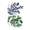
| ||||||||
|---|---|---|---|---|---|---|---|---|---|
| 1 |
| ||||||||
| Unit cell |
| ||||||||
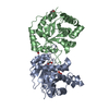
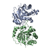
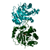
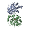
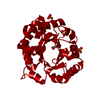
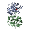
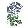
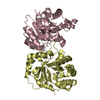
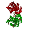
| Details | THE ASYMMETRIC UNIT CONSISTS OF A DIMER. THE TWO MOLECULES HAVE BEEN ASSIGNED CHAIN INDICATORS *A* AND *B*. |
-Components
| #1: Protein | Mass: 26865.832 Da / Num. of mol.: 2 Source method: isolated from a genetically manipulated source Source: (gene. exp.) #2: Chemical | ChemComp-2PG / | 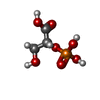  Mass: 186.057 Da / Num. of mol.: 1 / Source method: obtained synthetically / Formula: C3H7O7P Mass: 186.057 Da / Num. of mol.: 1 / Source method: obtained synthetically / Formula: C3H7O7P#3: Water | ChemComp-HOH / |  Mass: 18.015 Da / Num. of mol.: 130 / Source method: isolated from a natural source / Formula: H2O Mass: 18.015 Da / Num. of mol.: 130 / Source method: isolated from a natural source / Formula: H2OCompound details | SECONDARY STRUCTURE SPECIFICAT | |
|---|
-Experimental details
-Experiment
| Experiment | Method: X-RAY DIFFRACTION |
|---|
- Sample preparation
| Crystal | Density Matthews: 2.38 Å3/Da / Density % sol: 48.31 % | |||||||||||||||||||||||||||||||||||
|---|---|---|---|---|---|---|---|---|---|---|---|---|---|---|---|---|---|---|---|---|---|---|---|---|---|---|---|---|---|---|---|---|---|---|---|---|
| Crystal grow | Details: THE CRYSTALS USED FOR THIS STRUCTURE DETERMINATION WERE GROWN IN THE PRESENCE OF 2.4M AMMONIUM SULFATE (SEE PROTEIN DATA BANK ENTRIES 2TIM AND 5TIM), BUT BEFORE DATA COLLECTION THESE ...Details: THE CRYSTALS USED FOR THIS STRUCTURE DETERMINATION WERE GROWN IN THE PRESENCE OF 2.4M AMMONIUM SULFATE (SEE PROTEIN DATA BANK ENTRIES 2TIM AND 5TIM), BUT BEFORE DATA COLLECTION THESE CRYSTALS WERE TRANSFERRED TO A MOTHER LIQUOR WITHOUT SULFATE CONTAINING 30MM 2-PHOSPHOGLYCERATE. THE ACTIVE SITE OF CHAIN *A* ("OPEN"-CONFORMATION) HAS NO BOUND 2-PHOSPHOGLYCERATE. THE ACTIVE SITE OF CHAIN *B* ("CLOSED"-CONFORMATION) HAS A BOUND 2-PHOSPHOGLYCERATE. | |||||||||||||||||||||||||||||||||||
| Crystal grow | *PLUS pH: 7 / Method: vapor diffusion, hanging drop / Details: took 19 from original paper | |||||||||||||||||||||||||||||||||||
| Components of the solutions | *PLUS
|
-Data collection
| Reflection | *PLUS Highest resolution: 2.4 Å / Lowest resolution: 15 Å / Num. all: 17456 / % possible obs: 84.1 % / Rmerge(I) obs: 0.058 |
|---|
- Processing
| Software | Name: TNT / Classification: refinement | ||||||||||||||||||||||||||||||
|---|---|---|---|---|---|---|---|---|---|---|---|---|---|---|---|---|---|---|---|---|---|---|---|---|---|---|---|---|---|---|---|
| Refinement | Resolution: 2.4→15 Å /
| ||||||||||||||||||||||||||||||
| Refinement step | Cycle: LAST / Resolution: 2.4→15 Å
| ||||||||||||||||||||||||||||||
| Refine LS restraints |
| ||||||||||||||||||||||||||||||
| Software | *PLUS Name: TNT / Classification: refinement | ||||||||||||||||||||||||||||||
| Refinement | *PLUS Rfactor obs: 0.149 | ||||||||||||||||||||||||||||||
| Solvent computation | *PLUS | ||||||||||||||||||||||||||||||
| Displacement parameters | *PLUS | ||||||||||||||||||||||||||||||
| Refine LS restraints | *PLUS
|
