 Movie
Movie Controller

[English] 日本語
 Yorodumi
- PDB-2oma: Crystallographic analysis of a chemically modified triosephosphat... -
+ Open data

- Basic information
| Entry | Database: PDB / ID: 2oma | ||||||
|---|---|---|---|---|---|---|---|
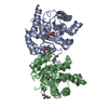
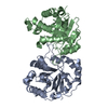
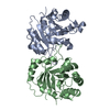
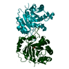
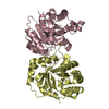
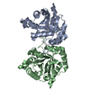
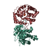
| Title | Crystallographic analysis of a chemically modified triosephosphate isomerase from Trypanosoma cruzi with dithiobenzylamine (DTBA) | ||||||
 Components Components | Triosephosphate isomerase | ||||||
 Keywords Keywords | ISOMERASE / Triosephosphate isomerase / Trypanosoma cruzi / protein interfaces / dithiobisbenzylamine | ||||||
| Function / homology |  Function and homology information Function and homology informationglycosome / triose-phosphate isomerase / triose-phosphate isomerase activity / glyceraldehyde-3-phosphate biosynthetic process / glycerol catabolic process / glycolytic process / gluconeogenesis / cytosol Similarity search - Function | ||||||
| Biological species |  | ||||||
| Method |  X-RAY DIFFRACTION / MOLECULAR REPLACEMENT / Resolution: 2.15 Å X-RAY DIFFRACTION / MOLECULAR REPLACEMENT / Resolution: 2.15 Å | ||||||
 Authors Authors | Rodriguez-Romero, A. / Gomez-Puyou, A. | ||||||
 Citation Citation | Journal: PLoS Negl Trop Dis / Year: 2007 Title: Perturbation of the Dimer Interface of Triosephosphate Isomerase and its Effect on Trypanosoma cruzi. Authors: Olivares-Illana, V. / Rodriguez-Romero, A. / Becker, I. / Berzunza, M. / Garcia, J. / Perez-Montfort, R. / Cabrera, N. / Lopez-Calahorra, F. / de Gomez-Puyou, M.T. / Gomez-Puyou, A. #1: Journal: J.Mol.Biol. / Year: 2004Title: Inactivation of triosephosphate isomerase from trypanosoma cruzi by an agent AN AGENT THAT PERTURBS ITS DIMER INTERFACE. Authors: Tellez-Valencia, A. / Olivares-Illana, V. / Hernandez-Santoyo, A. / Perez-Montfort, R. / Costas, M. / Rodriguez-Romero, A. / Lopez-Calahorra, F. / Tuena de Gomez-Puyou, M. / Gomez-Puyou, A. | ||||||
| History |
|
- Structure visualization
| Structure viewer | Molecule: MolmilJmol/JSmol |
|---|
- Downloads & links
-Download
| PDBx/mmCIF format | 2oma.cif.gz | 116.2 KB | Display | PDBx/mmCIF format |
|---|---|---|---|---|
| PDB format | pdb2oma.ent.gz | 89.2 KB | Display | PDB format |
| PDBx/mmJSON format | 2oma.json.gz | Tree view | PDBx/mmJSON format | |
| Others |  Other downloads Other downloads |
-Validation report
| Arichive directory | https://data.pdbj.org/pub/pdb/validation_reports/om/2omaftp://data.pdbj.org/pub/pdb/validation_reports/om/2oma | HTTPS FTP |
|---|
-Related structure data
| Related structure data |  1tcdS S: Starting model for refinement |
|---|---|
| Similar structure data |


-Links
 PDBj
PDBj
- Assembly
| Deposited unit | 
| ||||||||
|---|---|---|---|---|---|---|---|---|---|
| 1 |
| ||||||||
| Unit cell |
| ||||||||
| Details | The biological assembly is a dimer |
-Components
| #1: Protein | Mass: 27352.418 Da / Num. of mol.: 2 Source method: isolated from a genetically manipulated source Source: (gene. exp.)  #2: Chemical | ChemComp-SO4 /   Mass: 96.063 Da / Num. of mol.: 9 / Source method: obtained synthetically / Formula: SO4 Mass: 96.063 Da / Num. of mol.: 9 / Source method: obtained synthetically / Formula: SO4#3: Chemical |   Mass: 106.120 Da / Num. of mol.: 3 / Source method: obtained synthetically / Formula: C4H10O3 Mass: 106.120 Da / Num. of mol.: 3 / Source method: obtained synthetically / Formula: C4H10O3#4: Chemical | ChemComp-PGE / |   Mass: 150.173 Da / Num. of mol.: 1 / Source method: obtained synthetically / Formula: C6H14O4 Mass: 150.173 Da / Num. of mol.: 1 / Source method: obtained synthetically / Formula: C6H14O4#5: Water | ChemComp-HOH / |  Mass: 18.015 Da / Num. of mol.: 267 / Source method: isolated from a natural source / Formula: H2O Mass: 18.015 Da / Num. of mol.: 267 / Source method: isolated from a natural source / Formula: H2O |
|---|
-Experimental details
-Experiment
| Experiment | Method: X-RAY DIFFRACTION / Number of used crystals: 1 |
|---|
- Sample preparation
| Crystal | Density Matthews: 2.18 Å3/Da / Density % sol: 43.65 % |
|---|---|
| Crystal grow | Temperature: 291 K / pH: 7.5 Details: 5 MICROL OF THE PROTEIN SOLUTION WERE MIXED WITH 5 MICROL OF 2 % POLYETHYLENE GLYCOL 400, 0.1 M HEPES, 2.0M AMMONIUM SULFATE, PH 7.5, VAPOR DIFFUSION, HANGING DROP, TEMPERATURE 291K, PH 7.50. ...Details: 5 MICROL OF THE PROTEIN SOLUTION WERE MIXED WITH 5 MICROL OF 2 % POLYETHYLENE GLYCOL 400, 0.1 M HEPES, 2.0M AMMONIUM SULFATE, PH 7.5, VAPOR DIFFUSION, HANGING DROP, TEMPERATURE 291K, PH 7.50. CRYSTAL SOAKED IN DITHIOBENZYLAMINE |
-Data collection
| Diffraction | Mean temperature: 113 K |
|---|---|
| Diffraction source | Source: ROTATING ANODE / Type: RIGAKU RU200 / Wavelength: 1.5418 |
| Detector | Type: RIGAKU RAXIS IIC / Detector: IMAGE PLATE / Date: Mar 3, 2006 / Details: MIRRORS |
| Radiation | Protocol: SINGLE WAVELENGTH / Monochromatic (M) / Laue (L): M / Scattering type: x-ray |
| Radiation wavelength | Wavelength: 1.5418 Å / Relative weight: 1 |
| Reflection | Resolution: 2.15→41.45 Å / Num. obs: 23141 / % possible obs: 91 % / Redundancy: 3.4 % / Rmerge(I) obs: 0.093 / Net I/σ(I): 10.3 |
| Reflection shell | Resolution: 2.15→2.23 Å / Redundancy: 3.6 % / Rmerge(I) obs: 0.316 / Mean I/σ(I) obs: 3.8 / % possible all: 96.8 |
- Processing
| Software |
| ||||||||||||||||||||||||||||||||||||||||||||||||||||||||||||||||||||||||||||||||||||||||||||||||||||||||||||||||||||||||||||||||||||||||||||||||||||||||||||||||||||||||||
|---|---|---|---|---|---|---|---|---|---|---|---|---|---|---|---|---|---|---|---|---|---|---|---|---|---|---|---|---|---|---|---|---|---|---|---|---|---|---|---|---|---|---|---|---|---|---|---|---|---|---|---|---|---|---|---|---|---|---|---|---|---|---|---|---|---|---|---|---|---|---|---|---|---|---|---|---|---|---|---|---|---|---|---|---|---|---|---|---|---|---|---|---|---|---|---|---|---|---|---|---|---|---|---|---|---|---|---|---|---|---|---|---|---|---|---|---|---|---|---|---|---|---|---|---|---|---|---|---|---|---|---|---|---|---|---|---|---|---|---|---|---|---|---|---|---|---|---|---|---|---|---|---|---|---|---|---|---|---|---|---|---|---|---|---|---|---|---|---|---|---|---|
| Refinement | Method to determine structure: MOLECULAR REPLACEMENT Starting model: PDB ENTRY 1TCD Resolution: 2.15→41.45 Å / Cor.coef. Fo:Fc: 0.934 / Cor.coef. Fo:Fc free: 0.91 / SU B: 5.324 / SU ML: 0.137 / TLS residual ADP flag: LIKELY RESIDUAL / Cross valid method: THROUGHOUT / ESU R: 0.363 / ESU R Free: 0.241 / Stereochemistry target values: MAXIMUM LIKELIHOOD Details: HYDROGENS HAVE BEEN ADDED IN THE RIDING POSITIONS. ATOMS ARE ASSIGNED REDUCED OCCUPANCIES WHEN ELECTRON DENSITY IS WEAK OR ATOMS HAVE PARTIAL OCCUPANCY.
| ||||||||||||||||||||||||||||||||||||||||||||||||||||||||||||||||||||||||||||||||||||||||||||||||||||||||||||||||||||||||||||||||||||||||||||||||||||||||||||||||||||||||||
| Solvent computation | Ion probe radii: 0.8 Å / Shrinkage radii: 0.8 Å / VDW probe radii: 1.4 Å / Solvent model: MASK | ||||||||||||||||||||||||||||||||||||||||||||||||||||||||||||||||||||||||||||||||||||||||||||||||||||||||||||||||||||||||||||||||||||||||||||||||||||||||||||||||||||||||||
| Displacement parameters | Biso mean: 20.788 Å2
| ||||||||||||||||||||||||||||||||||||||||||||||||||||||||||||||||||||||||||||||||||||||||||||||||||||||||||||||||||||||||||||||||||||||||||||||||||||||||||||||||||||||||||
| Refinement step | Cycle: LAST / Resolution: 2.15→41.45 Å
| ||||||||||||||||||||||||||||||||||||||||||||||||||||||||||||||||||||||||||||||||||||||||||||||||||||||||||||||||||||||||||||||||||||||||||||||||||||||||||||||||||||||||||
| Refine LS restraints |
| ||||||||||||||||||||||||||||||||||||||||||||||||||||||||||||||||||||||||||||||||||||||||||||||||||||||||||||||||||||||||||||||||||||||||||||||||||||||||||||||||||||||||||
| LS refinement shell | Resolution: 2.15→2.206 Å / Total num. of bins used: 20
| ||||||||||||||||||||||||||||||||||||||||||||||||||||||||||||||||||||||||||||||||||||||||||||||||||||||||||||||||||||||||||||||||||||||||||||||||||||||||||||||||||||||||||
| Refinement TLS params. | Method: refined / Refine-ID: X-RAY DIFFRACTION
| ||||||||||||||||||||||||||||||||||||||||||||||||||||||||||||||||||||||||||||||||||||||||||||||||||||||||||||||||||||||||||||||||||||||||||||||||||||||||||||||||||||||||||
| Refinement TLS group |
|
