 Movie
Movie Controller

[English] 日本語
 Yorodumi
- PDB-5i3g: Structure-Function Studies on Role of Hydrophobic Clamping of a B... -
+ Open data

- Basic information
| Entry | Database: PDB / ID: 5i3g | ||||||
|---|---|---|---|---|---|---|---|
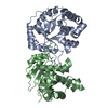
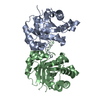
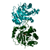
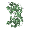
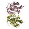
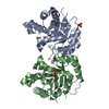
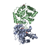
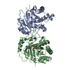
| Title | Structure-Function Studies on Role of Hydrophobic Clamping of a Basic Glutamate in Catalysis by Triosephosphate Isomerase | ||||||
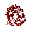
 Components Components | Triosephosphate isomerase, glycosomal | ||||||
 Keywords Keywords | ISOMERASE / Triosephosphate Isomerase / Catalysis / Hydrophobic Clamping | ||||||
| Function / homology |  Function and homology information Function and homology informationglycosome / triose-phosphate isomerase / triose-phosphate isomerase activity / glyceraldehyde-3-phosphate biosynthetic process / glycerol catabolic process / glycolytic process / gluconeogenesis / cytoplasm / cytosol Similarity search - Function | ||||||
| Biological species |  | ||||||
| Method |  X-RAY DIFFRACTION / MOLECULAR REPLACEMENT / Resolution: 1.96 Å X-RAY DIFFRACTION / MOLECULAR REPLACEMENT / Resolution: 1.96 Å | ||||||
 Authors Authors | Drake, E.J. / Gulick, A.M. / Richard, J.P. / Zhai, X. / Kim, K. / Reinhardt, C.J. | ||||||
 Citation Citation | Journal: Biochemistry / Year: 2016 Title: Structure-Function Studies of Hydrophobic Residues That Clamp a Basic Glutamate Side Chain during Catalysis by Triosephosphate Isomerase. Authors: Richard, J.P. / Amyes, T.L. / Malabanan, M.M. / Zhai, X. / Kim, K.J. / Reinhardt, C.J. / Wierenga, R.K. / Drake, E.J. / Gulick, A.M. | ||||||
| History |
|
- Structure visualization
| Structure viewer | Molecule: MolmilJmol/JSmol |
|---|
- Downloads & links
-Download
| PDBx/mmCIF format | 5i3g.cif.gz | 217.2 KB | Display | PDBx/mmCIF format |
|---|---|---|---|---|
| PDB format | pdb5i3g.ent.gz | 171.4 KB | Display | PDB format |
| PDBx/mmJSON format | 5i3g.json.gz | Tree view | PDBx/mmJSON format | |
| Others |  Other downloads Other downloads |
-Validation report
| Arichive directory | https://data.pdbj.org/pub/pdb/validation_reports/i3/5i3gftp://data.pdbj.org/pub/pdb/validation_reports/i3/5i3g | HTTPS FTP |
|---|
-Related structure data
| Related structure data |  5i3fC  5i3hC  5i3iC  5i3jC  5i3kC 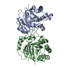 3timS C: citing same article ( S: Starting model for refinement |
|---|---|
| Similar structure data |


-Links
 PDBj
PDBj
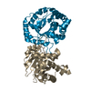
- Assembly
| Deposited unit | 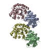
| ||||||||
|---|---|---|---|---|---|---|---|---|---|
| 1 | 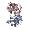
| ||||||||
| 2 | 
| ||||||||
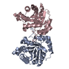
| Unit cell |
|
-Components
| #1: Protein | Mass: 26781.672 Da / Num. of mol.: 4 / Mutation: I172A,L232A Source method: isolated from a genetically manipulated source Source: (gene. exp.)  #2: Water | ChemComp-HOH / |  Mass: 18.015 Da / Num. of mol.: 1045 / Source method: isolated from a natural source / Formula: H2O Mass: 18.015 Da / Num. of mol.: 1045 / Source method: isolated from a natural source / Formula: H2O |
|---|
-Experimental details
-Experiment
| Experiment | Method: X-RAY DIFFRACTION / Number of used crystals: 1 |
|---|
- Sample preparation
| Crystal | Density Matthews: 2.29 Å3/Da / Density % sol: 46.22 % |
|---|---|
| Crystal grow | Temperature: 287 K / Method: vapor diffusion, hanging drop / pH: 7 Details: 15-25% Peg 8000, 50-100 mM potassium acetate, 100 mM BTP pH 7.0 |
-Data collection
| Diffraction | Mean temperature: 100 K |
|---|---|
| Diffraction source | Source: ROTATING ANODE / Type: RIGAKU / Wavelength: 1.5418 Å |
| Detector | Type: RIGAKU SATURN 944+ / Detector: CCD / Date: Jun 4, 2012 |
| Radiation | Protocol: SINGLE WAVELENGTH / Monochromatic (M) / Laue (L): M / Scattering type: x-ray |
| Radiation wavelength | Wavelength: 1.5418 Å / Relative weight: 1 |
| Reflection | Resolution: 1.96→29.03 Å / Num. obs: 67582 / % possible obs: 98.87 % / Redundancy: 2.7 % / Rmerge(I) obs: 0.097 / Net I/σ(I): 7 |
| Reflection shell | Resolution: 1.96→2.03 Å |
- Processing
| Software |
| |||||||||||||||||||||||||||||||||||||||||||||||||||||||||||||||||||||||||||||||||||||||||||||||||||||||||||||||||||||||||||||||||||||||||||||||||||||||||||||||||||||||||||||||
|---|---|---|---|---|---|---|---|---|---|---|---|---|---|---|---|---|---|---|---|---|---|---|---|---|---|---|---|---|---|---|---|---|---|---|---|---|---|---|---|---|---|---|---|---|---|---|---|---|---|---|---|---|---|---|---|---|---|---|---|---|---|---|---|---|---|---|---|---|---|---|---|---|---|---|---|---|---|---|---|---|---|---|---|---|---|---|---|---|---|---|---|---|---|---|---|---|---|---|---|---|---|---|---|---|---|---|---|---|---|---|---|---|---|---|---|---|---|---|---|---|---|---|---|---|---|---|---|---|---|---|---|---|---|---|---|---|---|---|---|---|---|---|---|---|---|---|---|---|---|---|---|---|---|---|---|---|---|---|---|---|---|---|---|---|---|---|---|---|---|---|---|---|---|---|---|---|
| Refinement | Method to determine structure: MOLECULAR REPLACEMENT Starting model: 3TIM Resolution: 1.96→29.03 Å / SU ML: 0.21 / Cross valid method: FREE R-VALUE / σ(F): 2.01 / Phase error: 26.13
| |||||||||||||||||||||||||||||||||||||||||||||||||||||||||||||||||||||||||||||||||||||||||||||||||||||||||||||||||||||||||||||||||||||||||||||||||||||||||||||||||||||||||||||||
| Solvent computation | Shrinkage radii: 0.9 Å / VDW probe radii: 1.11 Å | |||||||||||||||||||||||||||||||||||||||||||||||||||||||||||||||||||||||||||||||||||||||||||||||||||||||||||||||||||||||||||||||||||||||||||||||||||||||||||||||||||||||||||||||
| Refinement step | Cycle: LAST / Resolution: 1.96→29.03 Å
| |||||||||||||||||||||||||||||||||||||||||||||||||||||||||||||||||||||||||||||||||||||||||||||||||||||||||||||||||||||||||||||||||||||||||||||||||||||||||||||||||||||||||||||||
| Refine LS restraints |
| |||||||||||||||||||||||||||||||||||||||||||||||||||||||||||||||||||||||||||||||||||||||||||||||||||||||||||||||||||||||||||||||||||||||||||||||||||||||||||||||||||||||||||||||
| LS refinement shell |
|
