 Movie
Movie Controller


+ Open data

- Basic information
| Entry | Database: PDB / ID: 1nf0 | ||||||
|---|---|---|---|---|---|---|---|
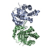
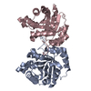
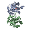
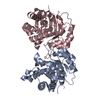
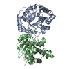
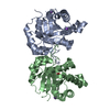
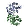
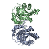
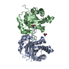
| Title | Triosephosphate Isomerase in Complex with DHAP | ||||||
 Components Components | triosephosphate isomerase | ||||||
 Keywords Keywords | ISOMERASE / yeast / triosephosphate isomerase / DHAP / dihydroxyacetone phosphate / michaelis complex | ||||||
| Function / homology |  Function and homology information Function and homology informationGluconeogenesis / Glycolysis / triose-phosphate isomerase / triose-phosphate isomerase activity / glyceraldehyde-3-phosphate biosynthetic process / glycerol catabolic process / gluconeogenesis / glycolytic process / mitochondrion / plasma membrane ...Gluconeogenesis / Glycolysis / triose-phosphate isomerase / triose-phosphate isomerase activity / glyceraldehyde-3-phosphate biosynthetic process / glycerol catabolic process / gluconeogenesis / glycolytic process / mitochondrion / plasma membrane / cytosol / cytoplasm Similarity search - Function | ||||||
| Biological species |  | ||||||
| Method |  X-RAY DIFFRACTION / SYNCHROTRON / COMO / Resolution: 1.6 Å X-RAY DIFFRACTION / SYNCHROTRON / COMO / Resolution: 1.6 Å | ||||||
 Authors Authors | Jogl, G. / Rozovsky, S. / McDermott, A.E. / Tong, L. | ||||||
 Citation Citation | Journal: Proc.Natl.Acad.Sci.USA / Year: 2003 Title: Optimal alignment for enzymatic proton transfer: Structure of the Michaelis complex of triosephosphate isomerase at 1.2-A resolution Authors: Jogl, G. / Rozovsky, S. / McDermott, A.E. / Tong, L. | ||||||
| History |
|
- Structure visualization
| Structure viewer | Molecule: MolmilJmol/JSmol |
|---|
- Downloads & links
-Download
| PDBx/mmCIF format | 1nf0.cif.gz | 118.6 KB | Display | PDBx/mmCIF format |
|---|---|---|---|---|
| PDB format | pdb1nf0.ent.gz | 90.8 KB | Display | PDB format |
| PDBx/mmJSON format | 1nf0.json.gz | Tree view | PDBx/mmJSON format | |
| Others |  Other downloads Other downloads |
-Validation report
| Arichive directory | https://data.pdbj.org/pub/pdb/validation_reports/nf/1nf0ftp://data.pdbj.org/pub/pdb/validation_reports/nf/1nf0 | HTTPS FTP |
|---|
-Related structure data
| Related structure data | 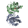 1neyC 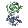 1i45S S: Starting model for refinement C: citing same article ( |
|---|---|
| Similar structure data |


-Links
 PDBj
PDBj
- Assembly
| Deposited unit | 
| ||||||||||
|---|---|---|---|---|---|---|---|---|---|---|---|
| 1 |
| ||||||||||
| Unit cell |
|
-Components
| #1: Protein | Mass: 26652.146 Da / Num. of mol.: 2 / Mutation: W90Y, W157F, W168(FTR) Source method: isolated from a genetically manipulated source Source: (gene. exp.) Gene: TPI1 / Plasmid: PKK223-3 / Production host:  #2: Chemical |   Mass: 170.058 Da / Num. of mol.: 2 / Source method: obtained synthetically / Formula: C3H7O6P Mass: 170.058 Da / Num. of mol.: 2 / Source method: obtained synthetically / Formula: C3H7O6P#3: Water | ChemComp-HOH / |  Mass: 18.015 Da / Num. of mol.: 419 / Source method: isolated from a natural source / Formula: H2O Mass: 18.015 Da / Num. of mol.: 419 / Source method: isolated from a natural source / Formula: H2O |
|---|
-Experimental details
-Experiment
| Experiment | Method: X-RAY DIFFRACTION / Number of used crystals: 1 |
|---|
- Sample preparation
| Crystal | Density Matthews: 1.91 Å3/Da / Density % sol: 44.43 % | ||||||||||||||||||||||||||||||||||||||||||
|---|---|---|---|---|---|---|---|---|---|---|---|---|---|---|---|---|---|---|---|---|---|---|---|---|---|---|---|---|---|---|---|---|---|---|---|---|---|---|---|---|---|---|---|
| Crystal grow | Temperature: 277 K / Method: evaporation, recrystallization / pH: 6.8 Details: 50mM TRIS, 50mM NaCl,20% PEG 4000,30mM DHAP, pH 6.8, EVAPORATION, RECRYSTALLIZATION, temperature 277K | ||||||||||||||||||||||||||||||||||||||||||
| Crystal grow | *PLUS Method: batch method / Details: Rozovsky, S., (2001) J. Mol. Biol., 310, 271. | ||||||||||||||||||||||||||||||||||||||||||
| Components of the solutions | *PLUS
|
-Data collection
| Diffraction | Mean temperature: 100 K |
|---|---|
| Diffraction source | Source: SYNCHROTRON / Site: CHESS  / Beamline: A1 / Wavelength: 0.928 Å / Beamline: A1 / Wavelength: 0.928 Å |
| Detector | Type: ADSC QUANTUM 4 / Detector: CCD / Date: Jan 4, 2001 |
| Radiation | Monochromator: NULL / Protocol: SINGLE WAVELENGTH / Monochromatic (M) / Laue (L): M / Scattering type: x-ray |
| Radiation wavelength | Wavelength: 0.928 Å / Relative weight: 1 |
| Reflection | Resolution: 1.6→40 Å / Num. all: 62255 / Num. obs: 62255 / % possible obs: 97.7 % / Observed criterion σ(F): 0 / Observed criterion σ(I): 0 |
| Reflection shell | Resolution: 1.6→1.66 Å / % possible all: 95.5 |
| Reflection | *PLUS Highest resolution: 1.6 Å / Num. measured all: 231083 / Rmerge(I) obs: 0.065 |
- Processing
| Software |
| |||||||||||||||||||||||||
|---|---|---|---|---|---|---|---|---|---|---|---|---|---|---|---|---|---|---|---|---|---|---|---|---|---|---|
| Refinement | Method to determine structure: COMO Starting model: pdb entry 1I45 Resolution: 1.6→40 Å / Num. parameters: 17233 / Num. restraintsaints: 16005 / Cross valid method: FREE R / σ(F): 0 / Stereochemistry target values: ENGH & HUBER Details: ANISOTROPIC SCALING APPLIED BY THE METHOD OF PARKIN, MOEZZI & HOPE, J.APPL.CRYST. 28(1995) 53-56
| |||||||||||||||||||||||||
| Refine analyze | Num. disordered residues: 18 / Occupancy sum hydrogen: 0 / Occupancy sum non hydrogen: 4194.32 | |||||||||||||||||||||||||
| Refinement step | Cycle: LAST / Resolution: 1.6→40 Å
| |||||||||||||||||||||||||
| Refine LS restraints |
| |||||||||||||||||||||||||
| Software | *PLUS Name: SHELXL / Version: 97 / Classification: refinement | |||||||||||||||||||||||||
| Refinement | *PLUS Highest resolution: 1.6 Å / Lowest resolution: 30 Å | |||||||||||||||||||||||||
| Solvent computation | *PLUS | |||||||||||||||||||||||||
| Displacement parameters | *PLUS |
