 Movie
Movie Controller

[English] 日本語
 Yorodumi
- PDB-2y61: Crystal structure of Leishmanial E65Q-TIM complexed with S-Glycid... -
+ Open data

- Basic information
| Entry | Database: PDB / ID: 2y61 | |||||||||
|---|---|---|---|---|---|---|---|---|---|---|
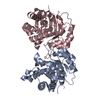
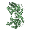
| Title | Crystal structure of Leishmanial E65Q-TIM complexed with S-Glycidol phosphate | |||||||||
 Components Components | TRIOSEPHOSPHATE ISOMERASE SYNONYM TRIOSE-PHOSPHATE ISOMERASE, TIM | |||||||||
 Keywords Keywords | ISOMERASE / FATTY ACID BIOSYNTHESIS / TRANSITION STATE ANALOGUE / GLYCOLYSIS / PENTOSE SHUNT / GLUCONEOGENESIS / ENZYME-LIGAND COMPLEX | |||||||||
| Function / homology |  Function and homology information Function and homology informationglycosome / triose-phosphate isomerase / triose-phosphate isomerase activity / glyceraldehyde-3-phosphate biosynthetic process / glycerol catabolic process / glycolytic process / gluconeogenesis / cytosol Similarity search - Function | |||||||||
| Biological species |   LEISHMANIA MEXICANA (eukaryote) LEISHMANIA MEXICANA (eukaryote) | |||||||||
| Method |  X-RAY DIFFRACTION / SYNCHROTRON / MOLECULAR REPLACEMENT / Resolution: 0.99 Å X-RAY DIFFRACTION / SYNCHROTRON / MOLECULAR REPLACEMENT / Resolution: 0.99 Å | |||||||||
 Authors Authors | Venkatesan, R. / Alahuhta, M. / Pihko, P.M. / Wierenga, R.K. | |||||||||
 Citation Citation | Journal: Protein Sci. / Year: 2011 Title: High resolution crystal structures of triosephosphate isomerase complexed with its suicide inhibitors: the conformational flexibility of the catalytic glutamate in its closed, liganded active site. Authors: Venkatesan, R. / Alahuhta, M. / Pihko, P.M. / Wierenga, R.K. | |||||||||
| History |
| |||||||||
| Remark 700 | SHEET DETERMINATION METHOD: DSSP THE SHEETS PRESENTED AS "AA" IN EACH CHAIN ON SHEET RECORDS BELOW ... SHEET DETERMINATION METHOD: DSSP THE SHEETS PRESENTED AS "AA" IN EACH CHAIN ON SHEET RECORDS BELOW IS ACTUALLY AN 8-STRANDED BARREL THIS IS REPRESENTED BY A 9-STRANDED SHEET IN WHICH THE FIRST AND LAST STRANDS ARE IDENTICAL. |
- Structure visualization
| Structure viewer | Molecule: MolmilJmol/JSmol |
|---|
- Downloads & links
-Download
| PDBx/mmCIF format | 2y61.cif.gz | 132.2 KB | Display | PDBx/mmCIF format |
|---|---|---|---|---|
| PDB format | pdb2y61.ent.gz | 102.4 KB | Display | PDB format |
| PDBx/mmJSON format | 2y61.json.gz | Tree view | PDBx/mmJSON format | |
| Others |  Other downloads Other downloads |
-Validation report
| Arichive directory | https://data.pdbj.org/pub/pdb/validation_reports/y6/2y61ftp://data.pdbj.org/pub/pdb/validation_reports/y6/2y61 | HTTPS FTP |
|---|
-Related structure data
| Related structure data | 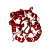 2y62C  2y63C  1n55S S: Starting model for refinement C: citing same article ( |
|---|---|
| Similar structure data |


-Links
 PDBj
PDBj
- Assembly
| Deposited unit | 
| ||||||||||||
|---|---|---|---|---|---|---|---|---|---|---|---|---|---|
| 1 | 
| ||||||||||||
| Unit cell |
| ||||||||||||
| Components on special symmetry positions |
|
-Components
| #1: Protein | Mass: 27208.236 Da / Num. of mol.: 1 / Mutation: E65Q Source method: isolated from a genetically manipulated source Source: (gene. exp.) LEISHMANIA MEXICANA (eukaryote) / Production host:  | ||||||||
|---|---|---|---|---|---|---|---|---|---|
| #2: Chemical | ChemComp-1GP /   Mass: 172.074 Da / Num. of mol.: 1 / Source method: obtained synthetically / Formula: C3H9O6P Mass: 172.074 Da / Num. of mol.: 1 / Source method: obtained synthetically / Formula: C3H9O6P | ||||||||
| #3: Chemical | ChemComp-G3P /   Mass: 172.074 Da / Num. of mol.: 1 / Source method: obtained synthetically / Formula: C3H9O6P Mass: 172.074 Da / Num. of mol.: 1 / Source method: obtained synthetically / Formula: C3H9O6P | ||||||||
| #4: Chemical |   Mass: 92.094 Da / Num. of mol.: 2 / Source method: obtained synthetically / Formula: C3H8O3 Mass: 92.094 Da / Num. of mol.: 2 / Source method: obtained synthetically / Formula: C3H8O3#5: Water | ChemComp-HOH / |  Mass: 18.015 Da / Num. of mol.: 417 / Source method: isolated from a natural source / Formula: H2O Mass: 18.015 Da / Num. of mol.: 417 / Source method: isolated from a natural source / Formula: H2OCompound details | ENGINEERED | Has protein modification | Y | Nonpolymer details | 1G3P, GOP: MICROHETEROGENEITY OBSERVED. S-GLYCIDOL PHOSPHATE BECAME GLYCEROL PHOSPHATE ESTER WITH ...1G3P, GOP: MICROHETER | |
-Experimental details
-Experiment
| Experiment | Method: X-RAY DIFFRACTION / Number of used crystals: 1 |
|---|
- Sample preparation
| Crystal | Density Matthews: 2.3 Å3/Da / Density % sol: 0.48 % / Description: NONE |
|---|---|
| Crystal grow | Temperature: 293 K / Method: vapor diffusion / pH: 5.5 Details: 21% PEG6000, 0.1 M SODIUM ACETATE PH 4.5-5.5, 1MM DTT, 1MM EDTA, 1MM NAN3 |
-Data collection
| Diffraction | Mean temperature: 100 K |
|---|---|
| Diffraction source | Source: SYNCHROTRON / Site: EMBL/DESY, HAMBURG  / Beamline: X12 / Wavelength: 0.8997 / Beamline: X12 / Wavelength: 0.8997 |
| Detector | Type: MARRESEARCH / Detector: CCD |
| Radiation | Protocol: SINGLE WAVELENGTH / Monochromatic (M) / Laue (L): M / Scattering type: x-ray |
| Radiation wavelength | Wavelength: 0.8997 Å / Relative weight: 1 |
| Reflection | Resolution: 0.99→10 Å / Num. obs: 140160 / % possible obs: 97.7 % / Observed criterion σ(I): 0 / Redundancy: 5.5 % / Rmerge(I) obs: 0.07 / Net I/σ(I): 21.4 |
| Reflection shell | Resolution: 0.99→1.02 Å / Redundancy: 4 % / Rmerge(I) obs: 0.44 / Mean I/σ(I) obs: 3.8 / % possible all: 95.3 |
- Processing
| Software |
| |||||||||||||||||||||||||||||||||
|---|---|---|---|---|---|---|---|---|---|---|---|---|---|---|---|---|---|---|---|---|---|---|---|---|---|---|---|---|---|---|---|---|---|---|
| Refinement | Method to determine structure: MOLECULAR REPLACEMENT Starting model: PDB ENTRY 1N55 Resolution: 0.99→10 Å / Num. parameters: 22147 / Num. restraintsaints: 27895 / Cross valid method: FREE R-VALUE / σ(F): 0
| |||||||||||||||||||||||||||||||||
| Refine analyze | Occupancy sum hydrogen: 1941.65 / Occupancy sum non hydrogen: 2322.88 | |||||||||||||||||||||||||||||||||
| Refinement step | Cycle: LAST / Resolution: 0.99→10 Å
| |||||||||||||||||||||||||||||||||
| Refine LS restraints |
|
