 Movie
Movie Controller

[English] 日本語
 Yorodumi
- PDB-4bxf: 60S ribosomal protein L27A histidine hydroxylase (MINA53 Y209C) i... -
+ Open data

- Basic information
| Entry | Database: PDB / ID: 4bxf | ||||||
|---|---|---|---|---|---|---|---|
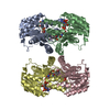
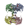
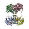
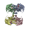
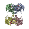
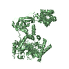
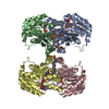
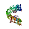
| Title | 60S ribosomal protein L27A histidine hydroxylase (MINA53 Y209C) in complex with MN(II), 2-oxoglutarate (2OG) and 60S ribosomal protein L27A (RPL27A G37C) peptide fragment | ||||||
 Components Components |
| ||||||
 Keywords Keywords | OXIDOREDUCTASE/TRANSLATION / OXIDOREDUCTASE-TRANSLATION COMPLEX / OXIDOREDUCTASE / NON-HEME / IRON-BINDING / DSBH / DIOXYGENASE / JMJC DOMAIN / RIBOSOME BIOGENESIS / NUCLEAR PROTEIN / BETA-HYDROXYLATION / TRANSCRIPTION AND EPIGENETIC REGULATION / SIGNALING | ||||||
| Function / homology |  Function and homology information Function and homology informationprotein-L-histidine (3S)-3-hydroxylase / peptidyl-histidine dioxygenase activity / histone H3K36 demethylase activity / histone H3K4 demethylase activity / histone demethylase activity / Protein hydroxylation / Peptide chain elongation / Selenocysteine synthesis / Formation of a pool of free 40S subunits / Eukaryotic Translation Termination ...protein-L-histidine (3S)-3-hydroxylase / peptidyl-histidine dioxygenase activity / histone H3K36 demethylase activity / histone H3K4 demethylase activity / histone demethylase activity / Protein hydroxylation / Peptide chain elongation / Selenocysteine synthesis / Formation of a pool of free 40S subunits / Eukaryotic Translation Termination / SRP-dependent cotranslational protein targeting to membrane / Response of EIF2AK4 (GCN2) to amino acid deficiency / Viral mRNA Translation / Nonsense Mediated Decay (NMD) independent of the Exon Junction Complex (EJC) / GTP hydrolysis and joining of the 60S ribosomal subunit / L13a-mediated translational silencing of Ceruloplasmin expression / Major pathway of rRNA processing in the nucleolus and cytosol / Nonsense Mediated Decay (NMD) enhanced by the Exon Junction Complex (EJC) / cytosolic ribosome / HDMs demethylate histones / Regulation of expression of SLITs and ROBOs / transcription corepressor activity / ribosome biogenesis / transcription regulator complex / cytosolic large ribosomal subunit / cytoplasmic translation / structural constituent of ribosome / translation / synapse / nucleolus / endoplasmic reticulum / RNA binding / nucleoplasm / metal ion binding / identical protein binding / membrane / cytoplasm / cytosol Similarity search - Function | ||||||
| Biological species |  HOMO SAPIENS (human) HOMO SAPIENS (human) | ||||||
| Method |  X-RAY DIFFRACTION / SYNCHROTRON / MOLECULAR REPLACEMENT / Resolution: 2.05 Å X-RAY DIFFRACTION / SYNCHROTRON / MOLECULAR REPLACEMENT / Resolution: 2.05 Å | ||||||
 Authors Authors | Chowdhury, R. / Schofield, C.J. | ||||||
 Citation Citation | Journal: Nature / Year: 2014 Title: Ribosomal oxygenases are structurally conserved from prokaryotes to humans. Authors: Chowdhury, R. / Sekirnik, R. / Brissett, N.C. / Krojer, T. / Ho, C.H. / Ng, S.S. / Clifton, I.J. / Ge, W. / Kershaw, N.J. / Fox, G.C. / Muniz, J.R.C. / Vollmar, M. / Phillips, C. / Pilka, E. ...Authors: Chowdhury, R. / Sekirnik, R. / Brissett, N.C. / Krojer, T. / Ho, C.H. / Ng, S.S. / Clifton, I.J. / Ge, W. / Kershaw, N.J. / Fox, G.C. / Muniz, J.R.C. / Vollmar, M. / Phillips, C. / Pilka, E.S. / Kavanagh, K.L. / von Delft, F. / Oppermann, U. / McDonough, M.A. / Doherty, A.J. / Schofield, C.J. #1: Journal: Nat.Chem.Biol. / Year: 2012 Title: Oxygenase-Catalyzed Ribosome Hydroxylation Occurs in Prokaryotes and Humans. Authors: Ge, W. / Wolf, A. / Feng, T. / Ho, C. / Sekirnik, R. / Zayer, A. / Granatino, N. / Cockman, M.E. / Loenarz, C. / Loik, N.D. / Hardy, A.P. / Claridge, T.D.W. / Hamed, R.B. / Chowdhury, R. / ...Authors: Ge, W. / Wolf, A. / Feng, T. / Ho, C. / Sekirnik, R. / Zayer, A. / Granatino, N. / Cockman, M.E. / Loenarz, C. / Loik, N.D. / Hardy, A.P. / Claridge, T.D.W. / Hamed, R.B. / Chowdhury, R. / Gong, L. / Robinson, C.V. / Trudgian, D.C. / Jiang, M. / Mackeen, M.M. / Mccullagh, J.S. / Gordiyenko, Y. / Thalhammer, A. / Yamamoto, A. / Yang, M. / Liu-Yi, P. / Zhang, Z. / Schmidt-Zachmann, M. / Kessler, B.M. / Ratcliffe, P.J. / Preston, G.M. / Coleman, M.L. / Schofield, C.J. | ||||||
| History |
|
- Structure visualization
| Structure viewer | Molecule: MolmilJmol/JSmol |
|---|
- Downloads & links
-Download
| PDBx/mmCIF format | 4bxf.cif.gz | 190.4 KB | Display | PDBx/mmCIF format |
|---|---|---|---|---|
| PDB format | pdb4bxf.ent.gz | 147.9 KB | Display | PDB format |
| PDBx/mmJSON format | 4bxf.json.gz | Tree view | PDBx/mmJSON format | |
| Others |  Other downloads Other downloads |
-Validation report
| Arichive directory | https://data.pdbj.org/pub/pdb/validation_reports/bx/4bxfftp://data.pdbj.org/pub/pdb/validation_reports/bx/4bxf | HTTPS FTP |
|---|
-Related structure data
| Related structure data | 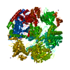 2xdvC 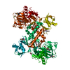 4bu2SC 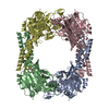 4ccjC 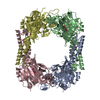 4cckC 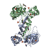 4cclC 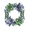 4ccmC 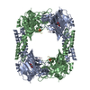 4ccnC 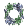 4ccoC 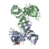 4cswC 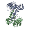 4cugC 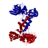 4litC 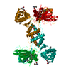 4liuC 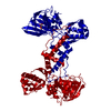 4livC C: citing same article ( S: Starting model for refinement |
|---|---|
| Similar structure data |


-Links
 PDBj
PDBj


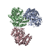
- Assembly
| Deposited unit | 
| ||||||||
|---|---|---|---|---|---|---|---|---|---|
| 1 |
| ||||||||
| Unit cell |
|
-Components
| #1: Protein | Mass: 50372.215 Da / Num. of mol.: 2 / Fragment: CATALYTIC DOMAIN, RESIDUES 26-465 / Mutation: YES Source method: isolated from a genetically manipulated source Source: (gene. exp.) HOMO SAPIENS (human) / Production host:  References: UniProt: Q8IUF8, Oxidoreductases; Acting on paired donors, with incorporation or reduction of molecular oxygen; With 2-oxoglutarate as one donor, and incorporation of one atom of oxygen into each donor #2: Protein/peptide | Mass: 2279.565 Da / Num. of mol.: 2 / Fragment: RESIDUES 32-50 / Mutation: YES / Source method: obtained synthetically / Source: (synth.) HOMO SAPIENS (human) / References: UniProt: P46776#3: Chemical |   Mass: 54.938 Da / Num. of mol.: 2 / Source method: obtained synthetically / Formula: Mn Mass: 54.938 Da / Num. of mol.: 2 / Source method: obtained synthetically / Formula: Mn#4: Chemical |   Mass: 146.098 Da / Num. of mol.: 2 / Source method: obtained synthetically / Formula: C5H6O5 Mass: 146.098 Da / Num. of mol.: 2 / Source method: obtained synthetically / Formula: C5H6O5#5: Water | ChemComp-HOH / |  Mass: 18.015 Da / Num. of mol.: 332 / Source method: isolated from a natural source / Formula: H2O Mass: 18.015 Da / Num. of mol.: 332 / Source method: isolated from a natural source / Formula: H2OHas protein modification | Y | |
|---|
-Experimental details
-Experiment
| Experiment | Method: X-RAY DIFFRACTION / Number of used crystals: 1 |
|---|
- Sample preparation
| Crystal | Density Matthews: 2.54 Å3/Da / Density % sol: 51.66 % / Description: NONE |
|---|---|
| Crystal grow | Temperature: 293 K / Method: vapor diffusion, sitting drop / pH: 8.5 Details: VAPOR DIFFUSION SITTING DROP. 0.1M BIS-TRIS PROPANE PH 8.5, 0.02M NA-K-PHOSPHATE, 20-22% (W/V) PEG 3350, 0.002 M MNCL2, TEMPERATURE 293K |
-Data collection
| Diffraction | Mean temperature: 100 K |
|---|---|
| Diffraction source | Source: SYNCHROTRON / Site: Diamond  / Beamline: I02 / Wavelength: 0.9795 / Beamline: I02 / Wavelength: 0.9795 |
| Detector | Type: DECTRIS PILATUS 6M / Detector: PIXEL / Date: Mar 16, 2013 / Details: MIRRORS |
| Radiation | Monochromator: SI 111 / Protocol: SINGLE WAVELENGTH / Monochromatic (M) / Laue (L): M / Scattering type: x-ray |
| Radiation wavelength | Wavelength: 0.9795 Å / Relative weight: 1 |
| Reflection | Resolution: 2.05→64.83 Å / Num. obs: 64784 / % possible obs: 98.2 % / Observed criterion σ(I): 2 / Redundancy: 3.3 % / Biso Wilson estimate: 31.2 Å2 / Rmerge(I) obs: 0.11 / Net I/σ(I): 7 |
| Reflection shell | Resolution: 2.05→2.16 Å / Redundancy: 3.4 % / Rmerge(I) obs: 0.9 / Mean I/σ(I) obs: 2 / % possible all: 97.3 |
- Processing
| Software |
| ||||||||||||||||||||||||||||||||||||||||||||||||||||||||||||
|---|---|---|---|---|---|---|---|---|---|---|---|---|---|---|---|---|---|---|---|---|---|---|---|---|---|---|---|---|---|---|---|---|---|---|---|---|---|---|---|---|---|---|---|---|---|---|---|---|---|---|---|---|---|---|---|---|---|---|---|---|---|
| Refinement | Method to determine structure: MOLECULAR REPLACEMENT Starting model: PDB ENTRY 4BU2 Resolution: 2.05→64.83 Å / Rfactor Rfree error: 0.002 / Data cutoff high absF: 298958.79 / Data cutoff low absF: 0 / Isotropic thermal model: RESTRAINED / Cross valid method: THROUGHOUT / σ(F): 0 / Stereochemistry target values: MAXIMUM LIKELIHOOD / Details: BULK SOLVENT MODEL USED
| ||||||||||||||||||||||||||||||||||||||||||||||||||||||||||||
| Solvent computation | Solvent model: FLAT MODEL / Bsol: 61.7167 Å2 / ksol: 0.4 e/Å3 | ||||||||||||||||||||||||||||||||||||||||||||||||||||||||||||
| Displacement parameters | Biso mean: 40.7 Å2
| ||||||||||||||||||||||||||||||||||||||||||||||||||||||||||||
| Refine analyze |
| ||||||||||||||||||||||||||||||||||||||||||||||||||||||||||||
| Refinement step | Cycle: LAST / Resolution: 2.05→64.83 Å
| ||||||||||||||||||||||||||||||||||||||||||||||||||||||||||||
| Refine LS restraints |
| ||||||||||||||||||||||||||||||||||||||||||||||||||||||||||||
| Refine LS restraints NCS | NCS model details: NONE | ||||||||||||||||||||||||||||||||||||||||||||||||||||||||||||
| LS refinement shell | Resolution: 2.05→2.16 Å / Rfactor Rfree error: 0.01 / Total num. of bins used: 7
| ||||||||||||||||||||||||||||||||||||||||||||||||||||||||||||
| Xplor file |
|
