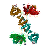
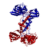
PDB-2xdv: Crystal Structure of the Catalytic Domain of FLJ14393 Method: X-RAY DIFFRACTION / Resolution: 2.57 Å
PDB-4bu2: 60S ribosomal protein L27A histidine hydroxylase (MINA53) in complex with Ni(II) and 2-oxoglutarate (2OG) Method: X-RAY DIFFRACTION / Resolution: 2.78 Å
PDB-4bxf: 60S ribosomal protein L27A histidine hydroxylase (MINA53 Y209C) in complex with MN(II), 2-oxoglutarate (2OG) and 60S ribosomal protein L27A (RPL27A G37C) peptide fragment Method: X-RAY DIFFRACTION / Resolution: 2.05 Å
PDB-4ccj: 60S ribosomal protein L8 histidine hydroxylase (NO66) in apo form Method: X-RAY DIFFRACTION / Resolution: 2.15 Å
PDB-4cck: 60S ribosomal protein L8 histidine hydroxylase (NO66) in complex with Mn(II) and N-oxalylglycine (NOG) Method: X-RAY DIFFRACTION / Resolution: 2.15 Å
PDB-4ccl: X-Ray structure of E. coli ycfD Method: X-RAY DIFFRACTION / Resolution: 2.596 Å
PDB-4ccm: 60S ribosomal protein L8 histidine hydroxylase (NO66) in complex with Mn(II), N-oxalylglycine (NOG) and 60S ribosomal protein L8 (RPL8 G220C) peptide fragment (complex-1) Method: X-RAY DIFFRACTION / Resolution: 2.51 Å
PDB-4ccn: 60S ribosomal protein L8 histidine hydroxylase (NO66 L299C/C300S) in complex with Mn(II), N-oxalylglycine (NOG) and 60S ribosomal protein L8 (RPL8 G220C) peptide fragment (complex-2) Method: X-RAY DIFFRACTION / Resolution: 2.23 Å
PDB-4cco: 60S ribosomal protein L8 histidine hydroxylase (NO66 S373C) in complex with Mn(II), N-oxalylglycine (NOG) and 60S ribosomal protein L8 (RPL8 G214C) peptide fragment (complex-3) Method: X-RAY DIFFRACTION / Resolution: 2.3 Å
PDB-4csw: Rhodothermus marinus YCFD-like ribosomal protein L16 Arginyl hydroxylase Method: X-RAY DIFFRACTION / Resolution: 2.821 Å
PDB-4cug: Rhodothermus marinus YCFD-like ribosomal protein L16 Arginyl hydroxylase in complex substrate fragment Method: X-RAY DIFFRACTION / Resolution: 2.96 Å
PDB-4lit: Structure of YcfD a Ribosomal oxygenase from Escherichia coli in complex with Cobalt and 2-oxoglutarate. Method: X-RAY DIFFRACTION / Resolution: 2.4 Å
PDB-4liu: Structure of YcfD, a Ribosomal oxygenase from Escherichia coli. Method: X-RAY DIFFRACTION / Resolution: 2.7 Å
PDB-4liv: Structure of YcfD, a Ribosomal oxygenase from Escherichia coli in complex with Cobalt and succinic acid. Method: X-RAY DIFFRACTION / Resolution: 2.7 Å
In the structure databanks used in Yorodumi, some data are registered as the other names, "COVID-19 virus" and "2019-nCoV". Here are the details of the virus and the list of structure data.
Jan 31, 2019. EMDB accession codes are about to change! (news from PDBe EMDB page)
EMDB accession codes are about to change! (news from PDBe EMDB page)
The allocation of 4 digits for EMDB accession codes will soon come to an end. Whilst these codes will remain in use, new EMDB accession codes will include an additional digit and will expand incrementally as the available range of codes is exhausted. The current 4-digit format prefixed with “EMD-” (i.e. EMD-XXXX) will advance to a 5-digit format (i.e. EMD-XXXXX), and so on. It is currently estimated that the 4-digit codes will be depleted around Spring 2019, at which point the 5-digit format will come into force.
The EM Navigator/Yorodumi systems omit the EMD- prefix.
Related info.:Q: What is EMD? / ID/Accession-code notation in Yorodumi/EM Navigator
 Movie
Movie Controller
 Structure viewers
 About Yorodumi Papers


 Authors
 External links


 Keywords
 homo sapiens (human)
 rhodothermus marinus (bacteria)