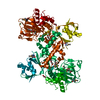
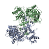
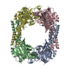
[50S ribosomal protein L16]-arginine 3-hydroxylase / 2-oxoglutarate-dependent dioxygenase activity / post-translational protein modification / ferrous iron binding / protein homodimerization activity Similarity search - Function
50S ribosomal protein L16 arginine hydroxylase; Chain A, Domain 2 / ROXA-like, winged helix / ROXA-like winged helix / JmjC domain-containing / JmjC domain / Malonyl-Coenzyme A Acyl Carrier Protein; domain 2 / Cupin / A domain family that is part of the cupin metalloenzyme superfamily. / JmjC domain / JmjC domain profile. ...50S ribosomal protein L16 arginine hydroxylase; Chain A, Domain 2 / ROXA-like, winged helix / ROXA-like winged helix / JmjC domain-containing / JmjC domain / Malonyl-Coenzyme A Acyl Carrier Protein; domain 2 / Cupin / A domain family that is part of the cupin metalloenzyme superfamily. / JmjC domain / JmjC domain profile. / Jelly Rolls / Sandwich / 3-Layer(aba) Sandwich / Mainly Beta / Alpha Beta Similarity search - Domain/homology
Mass: 46834.152 Da / Num. of mol.: 1 Source method: isolated from a genetically manipulated source Source: (gene. exp.) Escherichia coli (E. coli) / Strain: K-12 / Gene: b1128, JW1114, ycfD / Plasmid: pET28a / Production host: Escherichia coli (E. coli) / Strain (production host): B834s References: UniProt: P27431, Oxidoreductases; Acting on paired donors, with incorporation or reduction of molecular oxygen; With 2-oxoglutarate as one donor, and incorporation of one atom of oxygen into each donor
Monochromator: double crystal monochromator / Protocol: SAD / Monochromatic (M) / Laue (L): M / Scattering type: x-ray
Radiation wavelength
Wavelength: 0.9798 Å / Relative weight: 1
Reflection
Redundancy: 6.1 % / Av σ(I) over netI: 5.2 / Number: 106655 / Rsym value: 0.142 / D res high: 2.7 Å / D res low: 70.721 Å / Num. obs: 17365 / % possible obs: 99.9
Diffraction reflection shell
Highest resolution (Å)
Lowest resolution (Å)
% possible obs (%)
ID
Rmerge(I) obs
Rsym value
Redundancy
8.54
43.02
98.4
1
0.029
0.029
4.9
6.04
8.54
99.6
1
0.05
0.05
5.7
4.93
6.04
99.8
1
0.058
0.058
5.9
4.27
4.93
99.9
1
0.056
0.056
6.1
3.82
4.27
99.8
1
0.085
0.085
6.2
3.49
3.82
99.9
1
0.148
0.148
6.2
3.23
3.49
100
1
0.218
0.218
6.3
3.02
3.23
100
1
0.381
0.381
6.3
2.85
3.02
100
1
0.601
0.601
6.3
2.7
2.85
100
1
0.872
0.872
6.3
Reflection
Resolution: 2.7→70.721 Å / Num. all: 17365 / Num. obs: 17365 / % possible obs: 99.9 % / Redundancy: 6.1 % / Rsym value: 0.142 / Net I/σ(I): 11
Reflection shell
Resolution (Å)
Redundancy (%)
Rmerge(I) obs
Mean I/σ(I) obs
Rsym value
Diffraction-ID
% possible all
2.7-2.85
6.3
0.872
0.9
0.872
1
100
2.85-3.02
6.3
0.601
1.3
0.601
1
100
3.02-3.23
6.3
0.381
2
0.381
1
100
3.23-3.49
6.3
0.218
3.5
0.218
1
100
3.49-3.82
6.2
0.148
4.9
0.148
1
99.9
3.82-4.27
6.2
0.085
8.9
0.085
1
99.8
4.27-4.93
6.1
0.056
13
0.056
1
99.9
4.93-6.04
5.9
0.058
12.6
0.058
1
99.8
6.04-8.54
5.7
0.05
14.7
0.05
1
99.6
8.54-43.024
4.9
0.029
19
0.029
1
98.4
-
Phasing
Phasing
Method: SAD
Phasing MAD
D res high: 2.7 Å / D res low: 42.31 Å / FOM acentric: 0.233 / FOM centric: 0.057 / Reflection acentric: 14190 / Reflection centric: 3099
Phasing MAD set
ID
R cullis acentric
R cullis centric
Highest resolution (Å)
Lowest resolution (Å)
Reflection acentric
Reflection centric
ISO_1
0
0
2.7
42.31
14190
3099
ANO_1
0.894
0
2.7
42.31
14190
0
Phasing MAD set shell
ID
Resolution (Å)
R cullis acentric
R cullis centric
Reflection acentric
Reflection centric
ISO_1
11.63-42.31
0
0
120
143
ISO_1
8.39-11.63
0
0
249
148
ISO_1
6.89-8.39
0
0
345
155
ISO_1
5.99-6.89
0
0
418
151
ISO_1
5.37-5.99
0
0
482
151
ISO_1
4.91-5.37
0
0
548
159
ISO_1
4.55-4.91
0
0
594
155
ISO_1
4.26-4.55
0
0
643
158
ISO_1
4.01-4.26
0
0
691
156
ISO_1
3.81-4.01
0
0
732
145
ISO_1
3.63-3.81
0
0
777
165
ISO_1
3.48-3.63
0
0
819
157
ISO_1
3.35-3.48
0
0
854
154
ISO_1
3.22-3.35
0
0
874
160
ISO_1
3.12-3.22
0
0
923
154
ISO_1
3.02-3.12
0
0
959
154
ISO_1
2.93-3.02
0
0
1005
164
ISO_1
2.85-2.93
0
0
1020
154
ISO_1
2.77-2.85
0
0
1045
162
ISO_1
2.7-2.77
0
0
1092
154
ANO_1
11.63-42.31
0.336
0
120
0
ANO_1
8.39-11.63
0.32
0
249
0
ANO_1
6.89-8.39
0.398
0
345
0
ANO_1
5.99-6.89
0.47
0
418
0
ANO_1
5.37-5.99
0.629
0
482
0
ANO_1
4.91-5.37
0.631
0
548
0
ANO_1
4.55-4.91
0.71
0
594
0
ANO_1
4.26-4.55
0.789
0
643
0
ANO_1
4.01-4.26
0.835
0
691
0
ANO_1
3.81-4.01
0.892
0
732
0
ANO_1
3.63-3.81
0.906
0
777
0
ANO_1
3.48-3.63
0.949
0
819
0
ANO_1
3.35-3.48
0.959
0
854
0
ANO_1
3.22-3.35
0.968
0
874
0
ANO_1
3.12-3.22
0.981
0
923
0
ANO_1
3.02-3.12
0.993
0
959
0
ANO_1
2.93-3.02
0.993
0
1005
0
ANO_1
2.85-2.93
0.998
0
1020
0
ANO_1
2.77-2.85
1.001
0
1045
0
ANO_1
2.7-2.77
1.007
0
1092
0
Phasing MAD set site
ID
Cartn x (Å)
Cartn y (Å)
Cartn z (Å)
Atom type symbol
B iso
Occupancy
1
-61.238
-39.547
-17.06
SE
52.54
0.25
2
-55.873
-47.422
-29.466
SE
66.91
0.24
3
-38.127
-43.394
-48.366
SE
63.18
0.23
4
-57.022
-24.055
-1.988
SE
69.97
0.22
5
-73.244
-68.912
-11.569
SE
51.12
0.18
6
-5.46
-61.238
-5.699
SE
82.77
0.24
7
-71.434
-30.03
-23.417
SE
95.82
0.18
8
-53.116
-36.736
-24.849
SE
110.94
0.15
9
-14.146
-74.363
-25.349
SE
75.59
0.08
Phasing MAD shell
Resolution (Å)
FOM acentric
FOM centric
Reflection acentric
Reflection centric
11.63-42.31
0.623
0.079
120
143
8.39-11.63
0.597
0.092
249
148
6.89-8.39
0.573
0.094
345
155
5.99-6.89
0.522
0.084
418
151
5.37-5.99
0.458
0.094
482
151
4.91-5.37
0.462
0.065
548
159
4.55-4.91
0.419
0.054
594
155
4.26-4.55
0.37
0.05
643
158
4.01-4.26
0.344
0.047
691
156
3.81-4.01
0.286
0.04
732
145
3.63-3.81
0.26
0.04
777
165
3.48-3.63
0.211
0.047
819
157
3.35-3.48
0.195
0.042
854
154
3.22-3.35
0.154
0.041
874
160
3.12-3.22
0.135
0.043
923
154
3.02-3.12
0.112
0.045
959
154
2.93-3.02
0.102
0.051
1005
164
2.85-2.93
0.09
0.042
1020
154
2.77-2.85
0.078
0.053
1045
162
2.7-2.77
0.068
0.041
1092
154
-
Processing
Software
Name
Version
Classification
NB
MOSFLM
datareduction
SCALA
3.3.21
datascaling
SHARP
phasing
PHENIX
1.8.2_1309
refinement
PDB_EXTRACT
3.11
dataextraction
Refinement
Method to determine structure: SAD / Resolution: 2.7→43.024 Å / Occupancy max: 1 / Occupancy min: 0.49 / SU ML: 0.37 / σ(F): 1.01 / Phase error: 23.74 / Stereochemistry target values: ML
Rfactor
Num. reflection
% reflection
Rfree
0.2373
1590
5.07 %
Rwork
0.1966
-
-
obs
0.1986
31369
99.5 %
Solvent computation
Shrinkage radii: 0.9 Å / VDW probe radii: 1.11 Å / Solvent model: FLAT BULK SOLVENT MODEL
Displacement parameters
Biso mean: 40.9965 Å2
Refinement step
Cycle: LAST / Resolution: 2.7→43.024 Å
Protein
Nucleic acid
Ligand
Solvent
Total
Num. atoms
2875
0
16
113
3004
Refine LS restraints
Refine-ID
Type
Dev ideal
Number
X-RAY DIFFRACTION
f_bond_d
0.003
2989
X-RAY DIFFRACTION
f_angle_d
0.807
4074
X-RAY DIFFRACTION
f_dihedral_angle_d
15.2
1099
X-RAY DIFFRACTION
f_chiral_restr
0.06
417
X-RAY DIFFRACTION
f_plane_restr
0.003
545
LS refinement shell
Resolution (Å)
Rfactor Rfree
Num. reflection Rfree
Rfactor Rwork
Num. reflection Rwork
Refine-ID
% reflection obs (%)
2.7-2.7872
0.3857
162
0.2948
2690
X-RAY DIFFRACTION
99
2.7872-2.8868
0.3245
163
0.2753
2709
X-RAY DIFFRACTION
100
2.8868-3.0023
0.2944
140
0.27
2693
X-RAY DIFFRACTION
100
3.0023-3.1389
0.3033
174
0.2622
2711
X-RAY DIFFRACTION
100
3.1389-3.3043
0.2408
131
0.2173
2717
X-RAY DIFFRACTION
100
3.3043-3.5113
0.2194
142
0.1921
2696
X-RAY DIFFRACTION
100
3.5113-3.7822
0.2267
147
0.1898
2737
X-RAY DIFFRACTION
100
3.7822-4.1626
0.2246
136
0.1581
2710
X-RAY DIFFRACTION
100
4.1626-4.7643
0.168
154
0.1475
2692
X-RAY DIFFRACTION
100
4.7643-5.9999
0.2261
100
0.1732
2749
X-RAY DIFFRACTION
99
5.9999-43.0289
0.2329
141
0.1983
2675
X-RAY DIFFRACTION
98
Refinement TLS params.
Method: refined / Refine-ID: X-RAY DIFFRACTION
ID
L11 (°2)
L12 (°2)
L13 (°2)
L22 (°2)
L23 (°2)
L33 (°2)
S11 (Å °)
S12 (Å °)
S13 (Å °)
S21 (Å °)
S22 (Å °)
S23 (Å °)
S31 (Å °)
S32 (Å °)
S33 (Å °)
T11 (Å2)
T12 (Å2)
T13 (Å2)
T22 (Å2)
T23 (Å2)
T33 (Å2)
Origin x (Å)
Origin y (Å)
Origin z (Å)
1
8.4649
4.8263
-2.9061
6.4569
3.4402
8.0058
0.5197
-0.0361
-1.5878
0.4629
0.4337
-1.5751
0.695
1.3202
-0.3373
0.422
0.2808
-0.0227
0.6123
-0.0357
0.5195
22.2184
-18.418
31.1742
2
2.0525
1.654
-0.0359
1.8297
0.371
3.6234
-0.1184
-0.104
-0.5186
-0.0773
0.1213
-0.1254
0.4798
-0.3276
-0.0097
0.2137
-0.0176
0.0318
0.2149
-0.0121
0.2479
6.878
-16.1279
32.7988
3
3.9507
-0.3216
-1.5133
4.3546
0.7294
5.8428
0.2987
-0.0161
0.5722
-0.7135
0.2688
-0.4286
-1.204
0.2107
-0.3747
0.4289
-0.1473
-0.0006
0.4068
-0.0056
0.3214
22.8528
6.7613
30.4907
4
2.7482
-0.8239
-0.2051
1.0759
1.2178
3.393
0.1544
0.1338
-0.0031
-0.219
0.322
-0.7118
-0.4395
0.5838
-0.2975
0.295
-0.0639
0.0458
0.3331
-0.0288
0.2832
21.2648
-2.7954
27.3533
5
2.6185
0.2839
0.2857
3.0854
0.8033
3.8299
0.2355
-0.1202
0.2226
0.0909
-0.1036
0.1016
-0.4809
0.0003
-0.0758
0.2371
0.0148
0.0193
0.2115
0.0028
0.2064
9.7747
-2.3931
34.2245
6
2.5272
-1.9131
2.599
1.6971
-1.2706
4.0146
0.1498
-0.1044
0.1086
-0.0623
-0.0203
-0.0209
0.3718
0.1514
-0.146
0.3184
0.0395
0.0362
0.2505
0.0552
0.2587
-5.9257
0.429
-7.0225
7
6.6771
-2.266
3.4256
2.4221
-1.9138
2.0928
0.4664
1.0505
-0.2876
-0.6465
-0.2064
-0.1092
0.723
0.7256
-0.1914
0.5192
0.239
0.0292
0.5855
0.1007
0.3153
8.3666
-8.1959
3.071
8
3.0369
-2.1228
-0.6645
2.4343
-0.308
3.041
0.2498
0.2143
0.2593
-0.2313
-0.1207
-0.1689
-0.1978
-0.1323
-0.1569
0.205
0.0539
0.0445
0.2785
0.0069
0.2416
27.5488
-14.2774
7.4129
9
2.9065
-1.1196
-0.9207
4.4259
0.1359
2.0396
0.0248
0.0006
-0.2149
-0.0378
-0.1122
0.2876
0.1875
-0.0155
0.0912
0.2019
0.0405
0.0176
0.324
-0.0186
0.2335
27.1664
-20.6002
6.3548
Refinement TLS group
ID
Refine-ID
Refine TLS-ID
Selection details
1
X-RAY DIFFRACTION
1
(chainAandresid35:39)
2
X-RAY DIFFRACTION
2
(chainAandresid40:66)
3
X-RAY DIFFRACTION
3
(chainAandresid67:99)
4
X-RAY DIFFRACTION
4
(chainAandresid100:140)
5
X-RAY DIFFRACTION
5
(chainAandresid141:237)
6
X-RAY DIFFRACTION
6
(chainAandresid238:290)
7
X-RAY DIFFRACTION
7
(chainAandresid291:310)
8
X-RAY DIFFRACTION
8
(chainAandresid311:360)
9
X-RAY DIFFRACTION
9
(chainAandresid361:407)
+
About Yorodumi
-
News
-
Feb 9, 2022. New format data for meta-information of EMDB entries
New format data for meta-information of EMDB entries
Version 3 of the EMDB header file is now the official format.
The previous official version 1.9 will be removed from the archive.
In the structure databanks used in Yorodumi, some data are registered as the other names, "COVID-19 virus" and "2019-nCoV". Here are the details of the virus and the list of structure data.
Jan 31, 2019. EMDB accession codes are about to change! (news from PDBe EMDB page)
EMDB accession codes are about to change! (news from PDBe EMDB page)
The allocation of 4 digits for EMDB accession codes will soon come to an end. Whilst these codes will remain in use, new EMDB accession codes will include an additional digit and will expand incrementally as the available range of codes is exhausted. The current 4-digit format prefixed with “EMD-” (i.e. EMD-XXXX) will advance to a 5-digit format (i.e. EMD-XXXXX), and so on. It is currently estimated that the 4-digit codes will be depleted around Spring 2019, at which point the 5-digit format will come into force.
The EM Navigator/Yorodumi systems omit the EMD- prefix.
Related info.:Q: What is EMD? / ID/Accession-code notation in Yorodumi/EM Navigator
Yorodumi is a browser for structure data from EMDB, PDB, SASBDB, etc.
This page is also the successor to EM Navigator detail page, and also detail information page/front-end page for Omokage search.
The word "yorodu" (or yorozu) is an old Japanese word meaning "ten thousand". "mi" (miru) is to see.
Related info.:EMDB / PDB / SASBDB / Comparison of 3 databanks / Yorodumi Search / Aug 31, 2016. New EM Navigator & Yorodumi / Yorodumi Papers / Jmol/JSmol / Function and homology information / Changes in new EM Navigator and Yorodumi
 Movie
Movie Controller


 Open data
 Basic information
 Components
 Keywords
 Function and homology information
 X-RAY DIFFRACTION /
  Authors
 Citation
 Structure visualization
 Downloads & links
 Other downloads
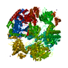


 PDBj
PDBj
 Assembly
 Mass: 163.215 Da / Num. of mol.: 1 / Source method: obtained synthetically / Formula: C7H17NO3 / Comment: pH buffer*YM
 Mass: 94.971 Da / Num. of mol.: 1 / Source method: obtained synthetically / Formula: PO4
 Mass: 18.015 Da / Num. of mol.: 113 / Source method: isolated from a natural source / Formula: H2O
 Sample preparation
 / Beamline: BM16 / Wavelength: 0.9798 Å
 Processing