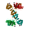
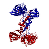
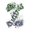
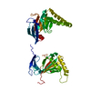
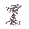
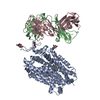
Crystal structure of Escherichia coli ribosomal oxygenase YcfD
Components
50S ribosomal protein L16 arginine hydroxylase
Keywords
OXIDOREDUCTASE / Structural Genomics / Montreal-Kingston Bacterial Structural Genomics Initiative / BSGI / jelly roll / cupin / beta-barrel / 2OG/Fe2+ dependent oxygenase / Ribosomal protein L-16
Function / homology
Function and homology information
[50S ribosomal protein L16]-arginine 3-hydroxylase / 2-oxoglutarate-dependent dioxygenase activity / post-translational protein modification / ferrous iron binding / protein homodimerization activity Similarity search - Function
50S ribosomal protein L16 arginine hydroxylase; Chain A, Domain 2 / ROXA-like, winged helix / ROXA-like winged helix / JmjC domain-containing / JmjC domain / Malonyl-Coenzyme A Acyl Carrier Protein; domain 2 / Cupin / A domain family that is part of the cupin metalloenzyme superfamily. / JmjC domain / JmjC domain profile. ...50S ribosomal protein L16 arginine hydroxylase; Chain A, Domain 2 / ROXA-like, winged helix / ROXA-like winged helix / JmjC domain-containing / JmjC domain / Malonyl-Coenzyme A Acyl Carrier Protein; domain 2 / Cupin / A domain family that is part of the cupin metalloenzyme superfamily. / JmjC domain / JmjC domain profile. / Jelly Rolls / Sandwich / 3-Layer(aba) Sandwich / Mainly Beta / Alpha Beta Similarity search - Domain/homology
Mass: 44333.980 Da / Num. of mol.: 1 / Mutation: E146A, K147A Source method: isolated from a genetically manipulated source Source: (gene. exp.) Escherichia coli (E. coli) / Strain: K12 / Gene: b1128, JW1114, ycfD / Plasmid: pET21b / Production host: Escherichia coli (E. coli) / Strain (production host): DL41 DE3 References: UniProt: P27431, Oxidoreductases; Acting on paired donors, with incorporation or reduction of molecular oxygen; With 2-oxoglutarate as one donor, and incorporation of one atom of oxygen into each donor
In the structure databanks used in Yorodumi, some data are registered as the other names, "COVID-19 virus" and "2019-nCoV". Here are the details of the virus and the list of structure data.
Jan 31, 2019. EMDB accession codes are about to change! (news from PDBe EMDB page)
EMDB accession codes are about to change! (news from PDBe EMDB page)
The allocation of 4 digits for EMDB accession codes will soon come to an end. Whilst these codes will remain in use, new EMDB accession codes will include an additional digit and will expand incrementally as the available range of codes is exhausted. The current 4-digit format prefixed with “EMD-” (i.e. EMD-XXXX) will advance to a 5-digit format (i.e. EMD-XXXXX), and so on. It is currently estimated that the 4-digit codes will be depleted around Spring 2019, at which point the 5-digit format will come into force.
The EM Navigator/Yorodumi systems omit the EMD- prefix.
Related info.:Q: What is EMD? / ID/Accession-code notation in Yorodumi/EM Navigator
Yorodumi is a browser for structure data from EMDB, PDB, SASBDB, etc.
This page is also the successor to EM Navigator detail page, and also detail information page/front-end page for Omokage search.
The word "yorodu" (or yorozu) is an old Japanese word meaning "ten thousand". "mi" (miru) is to see.
Related info.:EMDB / PDB / SASBDB / Comparison of 3 databanks / Yorodumi Search / Aug 31, 2016. New EM Navigator & Yorodumi / Yorodumi Papers / Jmol/JSmol / Function and homology information / Changes in new EM Navigator and Yorodumi
 Movie
Movie Controller


 Open data
 Basic information
 Components
 Keywords
 Function and homology information
 X-RAY DIFFRACTION /
  Authors
 Citation
 Structure visualization
 Downloads & links
 Other downloads
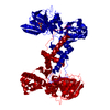

 PDBj
PDBj
 Assembly
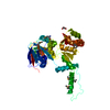

 Mass: 55.845 Da / Num. of mol.: 1 / Source method: obtained synthetically / Formula: Fe
 Mass: 92.094 Da / Num. of mol.: 1 / Source method: obtained synthetically / Formula: C3H8O3
 Mass: 18.015 Da / Num. of mol.: 51 / Source method: isolated from a natural source / Formula: H2O
 Sample preparation
 / Beamline: BL9-2 / Wavelength: 0.97913, 0.97927, 0.91837
 Processing