- PDB-2qkd: Crystal structure of tandem ZPR1 domains -
+
Open data
ID or keywords:
Loading...
-
Basic information
Entry
Database: PDB / ID: 2qkd
Title
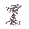
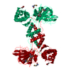
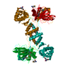
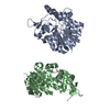
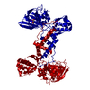
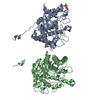
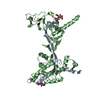
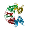
Crystal structure of tandem ZPR1 domains
Components
Zinc finger protein ZPR1
Keywords
SIGNALING PROTEIN / CELL CYCLE / HELICAL HAIRPINS / BETA HELIX / ANTI-PARRALLEL BETA SHEET / DOUBLE STRADED ANTI-PARALLEL BETA HELIX / METAL BINDING PROTEIN
Function / homology
Function and homology information
pre-mRNA catabolic process / DNA endoreduplication / positive regulation of growth / Gemini of Cajal bodies / Cajal body organization / trophectodermal cell proliferation / apoptotic process involved in development / negative regulation of motor neuron apoptotic process / regulation of myelination / inner cell mass cell proliferation ...pre-mRNA catabolic process / DNA endoreduplication / positive regulation of growth / Gemini of Cajal bodies / Cajal body organization / trophectodermal cell proliferation / apoptotic process involved in development / negative regulation of motor neuron apoptotic process / regulation of myelination / inner cell mass cell proliferation / axon development / spinal cord development / Cajal body / positive regulation of cell cycle / protein folding chaperone / translation initiation factor binding / RNA splicing / cellular response to epidermal growth factor stimulus / positive regulation of RNA splicing / positive regulation of protein import into nucleus / receptor tyrosine kinase binding / microtubule cytoskeleton organization / mRNA processing / growth cone / protein folding / perikaryon / DNA replication / axon / neuronal cell body / positive regulation of gene expression / perinuclear region of cytoplasm / nucleolus / zinc ion binding / nucleoplasm / nucleus / cytoplasm Similarity search - Function
ZPR1, zinc finger domain / ZPR1, A/B domain / Zinc finger, ZPR1-type / ZPR1 family / ZPR1, A/B domain / ZPR1, zinc finger domain / : / ZPR1 zinc-finger domain / ZPR1 jelly-roll domain / Duplicated domain in the epidermal growth factor- and elongation factor-1alpha-binding protein Zpr1. Also present in archaeal proteins. ...ZPR1, zinc finger domain / ZPR1, A/B domain / Zinc finger, ZPR1-type / ZPR1 family / ZPR1, A/B domain / ZPR1, zinc finger domain / : / ZPR1 zinc-finger domain / ZPR1 jelly-roll domain / Duplicated domain in the epidermal growth factor- and elongation factor-1alpha-binding protein Zpr1. Also present in archaeal proteins. / N-terminal domain of TfIIb / Single Sheet / Jelly Rolls / Sandwich / Mainly Beta Similarity search - Domain/homology
Monochromator: Si(111) / Protocol: SINGLE WAVELENGTH / Monochromatic (M) / Laue (L): M / Scattering type: x-ray
Radiation wavelength
Wavelength: 0.978715 Å / Relative weight: 1
Reflection
Resolution: 2→20 Å / Num. obs: 62931 / % possible obs: 98.8 % / Observed criterion σ(I): -3 / Redundancy: 3.7 % / Biso Wilson estimate: 42 Å2 / Rmerge(I) obs: 0.054 / Rsym value: 0.054 / Net I/σ(I): 29
Reflection shell
Resolution: 2→2.04 Å / Redundancy: 3.7 % / Rmerge(I) obs: 0.348 / Mean I/σ(I) obs: 3.16 / Num. unique all: 5305 / Rsym value: 0.348 / % possible all: 99.9
-
Processing
Software
Name
Version
Classification
REFMAC
5.2.0005
refinement
HKL-2000
datacollection
HKL-2000
datareduction
SCALEPACK
datascaling
SHARP
phasing
Refinement
Method to determine structure: SAD Starting model: heavy atom model Resolution: 2→6 Å / Cor.coef. Fo:Fc: 0.955 / Cor.coef. Fo:Fc free: 0.929 / SU B: 4.255 / SU ML: 0.122 / Isotropic thermal model: isotropic / Cross valid method: THROUGHOUT / σ(F): 2 / σ(I): 1 / ESU R: 0.19 / ESU R Free: 0.169 / Stereochemistry target values: MAXIMUM LIKELIHOOD Details: initial model generated by ARP/wARP was completed by manual building using O and refined in several iterative cycles usingARP/wARP and Refmac5.
Rfactor
Num. reflection
% reflection
Selection details
Rfree
0.24736
1569
5.1 %
RANDOM
Rwork
0.20617
-
-
-
all
0.218
-
-
-
obs
0.20835
29358
99.76 %
-
Solvent computation
Ion probe radii: 0.8 Å / Shrinkage radii: 0.8 Å / VDW probe radii: 1.2 Å / Solvent model: MASK
Displacement parameters
Biso mean: 42.135 Å2
Baniso -1
Baniso -2
Baniso -3
1-
1.27 Å2
0 Å2
0.73 Å2
2-
-
-0.78 Å2
0 Å2
3-
-
-
0.1 Å2
Refinement step
Cycle: LAST / Resolution: 2→6 Å
Protein
Nucleic acid
Ligand
Solvent
Total
Num. atoms
3021
0
2
278
3301
Refine LS restraints
Refine-ID
Type
Dev ideal
Dev ideal target
Number
X-RAY DIFFRACTION
r_bond_refined_d
0.01
0.022
3072
X-RAY DIFFRACTION
r_angle_refined_deg
1.265
1.971
4154
X-RAY DIFFRACTION
r_dihedral_angle_1_deg
6.168
5
382
X-RAY DIFFRACTION
r_dihedral_angle_2_deg
38.279
25.395
152
X-RAY DIFFRACTION
r_dihedral_angle_3_deg
14.115
15
559
X-RAY DIFFRACTION
r_dihedral_angle_4_deg
18.71
15
20
X-RAY DIFFRACTION
r_chiral_restr
0.082
0.2
468
X-RAY DIFFRACTION
r_gen_planes_refined
0.004
0.02
2330
X-RAY DIFFRACTION
r_nbd_refined
0.204
0.2
1190
X-RAY DIFFRACTION
r_nbtor_refined
0.298
0.2
2088
X-RAY DIFFRACTION
r_xyhbond_nbd_refined
0.144
0.2
252
X-RAY DIFFRACTION
r_symmetry_vdw_refined
0.191
0.2
99
X-RAY DIFFRACTION
r_symmetry_hbond_refined
0.135
0.2
24
X-RAY DIFFRACTION
r_mcbond_it
0.825
1.5
1994
X-RAY DIFFRACTION
r_mcangle_it
1.389
2
3111
X-RAY DIFFRACTION
r_scbond_it
1.979
3
1204
X-RAY DIFFRACTION
r_scangle_it
3.192
4.5
1043
LS refinement shell
Resolution: 2→2.06 Å / Rfactor Rfree error: 0.169 / Total num. of bins used: 20
Rfactor
Num. reflection
% reflection
Rfree
0.289
108
-
Rwork
0.219
2006
-
obs
-
29358
100 %
+
About Yorodumi
-
News
-
Feb 9, 2022. New format data for meta-information of EMDB entries
New format data for meta-information of EMDB entries
Version 3 of the EMDB header file is now the official format.
The previous official version 1.9 will be removed from the archive.
In the structure databanks used in Yorodumi, some data are registered as the other names, "COVID-19 virus" and "2019-nCoV". Here are the details of the virus and the list of structure data.
Jan 31, 2019. EMDB accession codes are about to change! (news from PDBe EMDB page)
EMDB accession codes are about to change! (news from PDBe EMDB page)
The allocation of 4 digits for EMDB accession codes will soon come to an end. Whilst these codes will remain in use, new EMDB accession codes will include an additional digit and will expand incrementally as the available range of codes is exhausted. The current 4-digit format prefixed with “EMD-” (i.e. EMD-XXXX) will advance to a 5-digit format (i.e. EMD-XXXXX), and so on. It is currently estimated that the 4-digit codes will be depleted around Spring 2019, at which point the 5-digit format will come into force.
The EM Navigator/Yorodumi systems omit the EMD- prefix.
Related info.:Q: What is EMD? / ID/Accession-code notation in Yorodumi/EM Navigator
Yorodumi is a browser for structure data from EMDB, PDB, SASBDB, etc.
This page is also the successor to EM Navigator detail page, and also detail information page/front-end page for Omokage search.
The word "yorodu" (or yorozu) is an old Japanese word meaning "ten thousand". "mi" (miru) is to see.
Related info.:EMDB / PDB / SASBDB / Comparison of 3 databanks / Yorodumi Search / Aug 31, 2016. New EM Navigator & Yorodumi / Yorodumi Papers / Jmol/JSmol / Function and homology information / Changes in new EM Navigator and Yorodumi
 Movie
Movie Controller


 Open data
 Basic information
 Components
 Keywords
 Function and homology information
 X-RAY DIFFRACTION /
  Authors
 Citation
 Structure visualization
 Downloads & links
 Other downloads
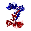
 PDBj
PDBj Assembly
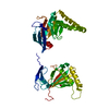


 Mass: 65.409 Da / Num. of mol.: 2 / Source method: obtained synthetically / Formula: Zn
 Mass: 46.025 Da / Num. of mol.: 1 / Source method: obtained synthetically / Formula: CH2O2
 Mass: 18.015 Da / Num. of mol.: 275 / Source method: isolated from a natural source / Formula: H2O
 Sample preparation
 / Beamline: X25 / Wavelength: 0.978715 Å
 Processing