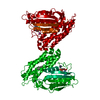
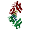
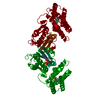
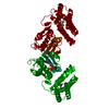
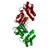
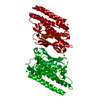
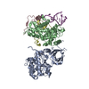
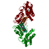
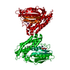
Entry Database : PDB / ID : 3tz4Title Crystal structure of branched-chain alpha-ketoacid dehydrogenase kinase/S-alpha-chloroisocaproate complex with ADP [3-methyl-2-oxobutanoate dehydrogenase [lipoamide]] kinase, mitochondrial Keywords / / / / / / / / / Function / homology Function Domain/homology Component
/ / / / / / / / / / / / / / / / / / / / / / / / / / / / / / / / / / / / / / / / / / / / / / / / / / / / / / / / / / / Biological species Rattus norvegicus (Norway rat)Method / / / Resolution : 2.25 Å Authors Tso, S.C. / Chuang, J.L. / Gui, W.J. / Wynn, R.M. / Li, J. / Chuang, D.T. Journal : Proc.Natl.Acad.Sci.USA / Year : 2013Title : Structure-based design and mechanisms of allosteric inhibitors for mitochondrial branched-chain alpha-ketoacid dehydrogenase kinase.Authors: Tso, S.C. / Qi, X. / Gui, W.J. / Chuang, J.L. / Morlock, L.K. / Wallace, A.L. / Ahmed, K. / Laxman, S. / Campeau, P.M. / Lee, B.H. / Hutson, S.M. / Tu, B.P. / Williams, N.S. / Tambar, U.K. / ... Authors : Tso, S.C. / Qi, X. / Gui, W.J. / Chuang, J.L. / Morlock, L.K. / Wallace, A.L. / Ahmed, K. / Laxman, S. / Campeau, P.M. / Lee, B.H. / Hutson, S.M. / Tu, B.P. / Williams, N.S. / Tambar, U.K. / Wynn, R.M. / Chuang, D.T. History Deposition Sep 27, 2011 Deposition site / Processing site Revision 1.0 Oct 3, 2012 Provider / Type Revision 1.1 May 29, 2013 Group Revision 1.2 Sep 18, 2019 Group / Database references / Category / citation_authorItem _citation.journal_volume / _citation.page_first ... _citation.journal_volume / _citation.page_first / _citation.page_last / _citation.pdbx_database_id_DOI / _citation.pdbx_database_id_PubMed / _citation.title Revision 1.3 Feb 28, 2024 Group / Database references / Derived calculationsCategory chem_comp_atom / chem_comp_bond ... chem_comp_atom / chem_comp_bond / database_2 / pdbx_struct_conn_angle / struct_conn / struct_ref_seq_dif Item _database_2.pdbx_DOI / _database_2.pdbx_database_accession ... _database_2.pdbx_DOI / _database_2.pdbx_database_accession / _pdbx_struct_conn_angle.ptnr1_auth_comp_id / _pdbx_struct_conn_angle.ptnr1_auth_seq_id / _pdbx_struct_conn_angle.ptnr1_label_asym_id / _pdbx_struct_conn_angle.ptnr1_label_atom_id / _pdbx_struct_conn_angle.ptnr1_label_comp_id / _pdbx_struct_conn_angle.ptnr1_label_seq_id / _pdbx_struct_conn_angle.ptnr3_auth_comp_id / _pdbx_struct_conn_angle.ptnr3_auth_seq_id / _pdbx_struct_conn_angle.ptnr3_label_asym_id / _pdbx_struct_conn_angle.ptnr3_label_atom_id / _pdbx_struct_conn_angle.ptnr3_label_comp_id / _pdbx_struct_conn_angle.ptnr3_label_seq_id / _pdbx_struct_conn_angle.value / _struct_conn.pdbx_dist_value / _struct_conn.ptnr1_auth_comp_id / _struct_conn.ptnr1_auth_seq_id / _struct_conn.ptnr1_label_asym_id / _struct_conn.ptnr1_label_atom_id / _struct_conn.ptnr1_label_comp_id / _struct_conn.ptnr1_label_seq_id / _struct_conn.ptnr2_auth_comp_id / _struct_conn.ptnr2_auth_seq_id / _struct_conn.ptnr2_label_asym_id / _struct_conn.ptnr2_label_atom_id / _struct_conn.ptnr2_label_comp_id / _struct_ref_seq_dif.details
Show all Show less
 Movie
Movie Controller

 Yorodumi
 Open data
 Basic information
 Components
 Keywords
 Function and homology information
 X-RAY DIFFRACTION /
  Authors
 Citation
 Structure visualization
 Downloads & links
 Other downloads


 PDBj
PDBj

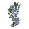
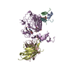
 Assembly
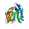


 Mass: 427.201 Da / Num. of mol.: 1 / Source method: obtained synthetically / Formula: C10H15N5O10P2 / Comment: ADP, energy-carrying molecule*YM
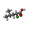
 Mass: 150.603 Da / Num. of mol.: 1 / Source method: obtained synthetically / Formula: C6H11ClO2
 Mass: 24.305 Da / Num. of mol.: 1 / Source method: obtained synthetically / Formula: Mg
 Mass: 39.098 Da / Num. of mol.: 1 / Source method: obtained synthetically / Formula: K
 Sample preparation
 / Beamline: 19-ID / Wavelength: 0.97864 Å
 Processing