 Movie
Movie Controller

[English] 日本語
 Yorodumi
- PDB-3tz0: Crystal structure of branched-chain alpha-ketoacid dehydrogenase ... -
+ Open data

- Basic information
| Entry | Database: PDB / ID: 3tz0 | ||||||
|---|---|---|---|---|---|---|---|
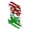
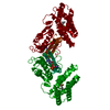
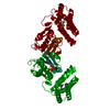
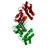
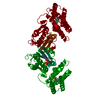
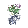
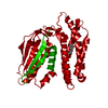
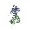
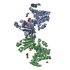
| Title | Crystal structure of branched-chain alpha-ketoacid dehydrogenase kinase/S-alpha-chloroisocaproate complex | ||||||
 Components Components | [3-methyl-2-oxobutanoate dehydrogenase [lipoamide]] kinase, mitochondrial | ||||||
 Keywords Keywords | TRANSFERASE/TRANSFERASE INHIBITOR / GHKL protein kinase / Allosteric kinase inhibitor / Branched-chain alpha-ketoacid / Branched-chain amino acids / Maple syrup urine disease / Diabetes and obesity / Bergerat nucleotide-binding fold / Protein kinase / TRANSFERASE-TRANSFERASE INHIBITOR complex | ||||||
| Function / homology |  Function and homology information Function and homology information[3-methyl-2-oxobutanoate dehydrogenase (acetyl-transferring)] kinase / [3-methyl-2-oxobutanoate dehydrogenase (acetyl-transferring)] kinase activity / Branched-chain amino acid catabolism / pyruvate dehydrogenase (acetyl-transferring) kinase activity / regulation of pyruvate decarboxylation to acetyl-CoA / L-isoleucine catabolic process / L-valine catabolic process / L-leucine catabolic process / oxoglutarate dehydrogenase complex / branched-chain amino acid catabolic process ...[3-methyl-2-oxobutanoate dehydrogenase (acetyl-transferring)] kinase / [3-methyl-2-oxobutanoate dehydrogenase (acetyl-transferring)] kinase activity / Branched-chain amino acid catabolism / pyruvate dehydrogenase (acetyl-transferring) kinase activity / regulation of pyruvate decarboxylation to acetyl-CoA / L-isoleucine catabolic process / L-valine catabolic process / L-leucine catabolic process / oxoglutarate dehydrogenase complex / branched-chain amino acid catabolic process / lipid biosynthetic process / protein serine/threonine phosphatase activity / regulation of glucose metabolic process / spermatogenesis / protein kinase activity / non-specific serine/threonine protein kinase / mitochondrial matrix / protein serine kinase activity / protein serine/threonine kinase activity / mitochondrion / ATP binding Similarity search - Function | ||||||
| Biological species |  | ||||||
| Method |  X-RAY DIFFRACTION / SYNCHROTRON / MOLECULAR REPLACEMENT / Resolution: 2.5 Å X-RAY DIFFRACTION / SYNCHROTRON / MOLECULAR REPLACEMENT / Resolution: 2.5 Å | ||||||
 Authors Authors | Tso, S.C. / Chuang, J.L. / Gui, W.J. / Wynn, R.M. / Li, J. / Chuang, D.T. | ||||||
 Citation Citation | Journal: Proc.Natl.Acad.Sci.USA / Year: 2013 Title: Structure-based design and mechanisms of allosteric inhibitors for mitochondrial branched-chain alpha-ketoacid dehydrogenase kinase. Authors: Tso, S.C. / Qi, X. / Gui, W.J. / Chuang, J.L. / Morlock, L.K. / Wallace, A.L. / Ahmed, K. / Laxman, S. / Campeau, P.M. / Lee, B.H. / Hutson, S.M. / Tu, B.P. / Williams, N.S. / Tambar, U.K. / ...Authors: Tso, S.C. / Qi, X. / Gui, W.J. / Chuang, J.L. / Morlock, L.K. / Wallace, A.L. / Ahmed, K. / Laxman, S. / Campeau, P.M. / Lee, B.H. / Hutson, S.M. / Tu, B.P. / Williams, N.S. / Tambar, U.K. / Wynn, R.M. / Chuang, D.T. | ||||||
| History |
|
- Structure visualization
| Structure viewer | Molecule: MolmilJmol/JSmol |
|---|
- Downloads & links
-Download
| PDBx/mmCIF format | 3tz0.cif.gz | 141.7 KB | Display | PDBx/mmCIF format |
|---|---|---|---|---|
| PDB format | pdb3tz0.ent.gz | 110.3 KB | Display | PDB format |
| PDBx/mmJSON format | 3tz0.json.gz | Tree view | PDBx/mmJSON format | |
| Others |  Other downloads Other downloads |
-Validation report
| Arichive directory | https://data.pdbj.org/pub/pdb/validation_reports/tz/3tz0ftp://data.pdbj.org/pub/pdb/validation_reports/tz/3tz0 | HTTPS FTP |
|---|
-Related structure data
| Related structure data |  3tz2C  3tz4C 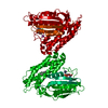 3tz5C  4dzyC  4h7qC  4h81C  4h85C C: citing same article ( |
|---|---|
| Similar structure data |

-Links
 PDBj
PDBj

- Assembly
| Deposited unit | 
| ||||||||
|---|---|---|---|---|---|---|---|---|---|
| 1 | 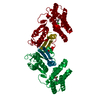
| ||||||||
| 2 |
| ||||||||
| Unit cell |
| ||||||||
| Details | THE AUTHORS PROVIDED BURIED SURFACE AREA IS 924.7 A2 |
-Components
| #1: Protein | [ Mass: 47374.246 Da / Num. of mol.: 1 Source method: isolated from a genetically manipulated source Source: (gene. exp.)  References: UniProt: Q00972, [3-methyl-2-oxobutanoate dehydrogenase (acetyl-transferring)] kinase |
|---|---|
| #2: Chemical | ChemComp-03H / (  Mass: 150.603 Da / Num. of mol.: 1 / Source method: obtained synthetically / Formula: C6H11ClO2 Mass: 150.603 Da / Num. of mol.: 1 / Source method: obtained synthetically / Formula: C6H11ClO2 |
| #3: Water | ChemComp-HOH /  Mass: 18.015 Da / Num. of mol.: 21 / Source method: isolated from a natural source / Formula: H2O Mass: 18.015 Da / Num. of mol.: 21 / Source method: isolated from a natural source / Formula: H2O |
-Experimental details
-Experiment
| Experiment | Method: X-RAY DIFFRACTION / Number of used crystals: 1 |
|---|
- Sample preparation
| Crystal | Density Matthews: 3.19 Å3/Da / Density % sol: 61.46 % |
|---|---|
| Crystal grow | Temperature: 289 K / Method: vapor diffusion, hanging drop / pH: 7.5 Details: 14% peg8000, 0.1 M Tris, pH8.5, 1.2 M NaCl, 125mM KCl, 150mM Arg-HCl,20mM MgCl2, 5% glycerol , pH 7.5, VAPOR DIFFUSION, HANGING DROP, temperature 289K |
-Data collection
| Diffraction | Mean temperature: 100 K |
|---|---|
| Diffraction source | Source: SYNCHROTRON / Site: APS  / Beamline: 19-ID / Wavelength: 0.97926 Å / Beamline: 19-ID / Wavelength: 0.97926 Å |
| Detector | Type: ADSC QUANTUM 315r / Detector: CCD / Date: Aug 31, 2011 / Details: mirrors |
| Radiation | Monochromator: double-crystal / Protocol: SINGLE WAVELENGTH / Monochromatic (M) / Laue (L): M / Scattering type: x-ray |
| Radiation wavelength | Wavelength: 0.97926 Å / Relative weight: 1 |
| Reflection | Resolution: 2.5→50 Å / Num. all: 21812 / Num. obs: 21439 / % possible obs: 98.3 % / Observed criterion σ(F): 2 / Observed criterion σ(I): 2 / Redundancy: 7.9 % / Rmerge(I) obs: 0.05 / Rsym value: 0.036 / Net I/σ(I): 36.9 |
| Reflection shell | Resolution: 2.5→2.54 Å / Redundancy: 6.6 % / Rmerge(I) obs: 0.711 / Mean I/σ(I) obs: 2.24 / Num. unique all: 1046 / Rsym value: 0.661 / % possible all: 98.2 |
- Processing
| Software |
| |||||||||||||||||||||||||||||||||||||||||||||||||||||||||||||||||||||||||||||||||||||||||||||||||||||||||
|---|---|---|---|---|---|---|---|---|---|---|---|---|---|---|---|---|---|---|---|---|---|---|---|---|---|---|---|---|---|---|---|---|---|---|---|---|---|---|---|---|---|---|---|---|---|---|---|---|---|---|---|---|---|---|---|---|---|---|---|---|---|---|---|---|---|---|---|---|---|---|---|---|---|---|---|---|---|---|---|---|---|---|---|---|---|---|---|---|---|---|---|---|---|---|---|---|---|---|---|---|---|---|---|---|---|---|
| Refinement | Method to determine structure: MOLECULAR REPLACEMENT / Resolution: 2.5→45.199 Å / SU ML: 0.7 / σ(F): 1.34 / Phase error: 23.68 / Stereochemistry target values: ML
| |||||||||||||||||||||||||||||||||||||||||||||||||||||||||||||||||||||||||||||||||||||||||||||||||||||||||
| Solvent computation | Shrinkage radii: 0.95 Å / VDW probe radii: 1.2 Å / Solvent model: FLAT BULK SOLVENT MODEL / Bsol: 63.642 Å2 / ksol: 0.373 e/Å3 | |||||||||||||||||||||||||||||||||||||||||||||||||||||||||||||||||||||||||||||||||||||||||||||||||||||||||
| Displacement parameters |
| |||||||||||||||||||||||||||||||||||||||||||||||||||||||||||||||||||||||||||||||||||||||||||||||||||||||||
| Refinement step | Cycle: LAST / Resolution: 2.5→45.199 Å
| |||||||||||||||||||||||||||||||||||||||||||||||||||||||||||||||||||||||||||||||||||||||||||||||||||||||||
| Refine LS restraints |
| |||||||||||||||||||||||||||||||||||||||||||||||||||||||||||||||||||||||||||||||||||||||||||||||||||||||||
| LS refinement shell |
| |||||||||||||||||||||||||||||||||||||||||||||||||||||||||||||||||||||||||||||||||||||||||||||||||||||||||
| Refinement TLS params. | Method: refined / Origin x: 3.4143 Å / Origin y: -30.72 Å / Origin z: 10.3519 Å
| |||||||||||||||||||||||||||||||||||||||||||||||||||||||||||||||||||||||||||||||||||||||||||||||||||||||||
| Refinement TLS group | Selection details: all |
