#1: Journal: Acta Crystallogr. Sect. F Struct. Biol. Cryst. Commun. Year: 2008 Title: Purification, crystallization and preliminary X-ray diffraction analysis of human chondroadherin. Authors: Pramhed, A. / Addis, L. / Tillgren, V. / Wenglen, C. / Heinegard, D. / Logan, D.T.
History
Deposition
Jul 3, 2016
Deposition site: PDBE / Processing site: PDBE
Revision 1.0
Dec 28, 2016
Provider: repository / Type: Initial release
Revision 1.1
Jan 11, 2017
Group: Database references
Revision 1.2
Jan 25, 2017
Group: Database references
Revision 1.3
Jan 17, 2018
Group: Data collection / Category: diffrn_source / Item: _diffrn_source.pdbx_synchrotron_site
Mass: 18.015 Da / Num. of mol.: 120 / Source method: isolated from a natural source / Formula: H2O
Has protein modification
Y
-
Experimental details
-
Experiment
Experiment
Method: X-RAY DIFFRACTION / Number of used crystals: 1
-
Sample preparation
Crystal
Density Matthews: 2.63 Å3/Da / Density % sol: 53.8 %
Crystal grow
Temperature: 293 K / Method: vapor diffusion / pH: 9.5 Details: 21% PEG 3350, 6% xylitol, 0.1 M glycine-NaOH pH 9.5-10.5, 0.2 M ammonium sulfate. Protein at 1.2 mg/ml in 20 mM Tris pH 7.0, 200 mM NaCl. Mixed in 1:1 ratio with reservoir.
-
Data collection
Diffraction
Mean temperature: 100 K
Diffraction source
Source: SYNCHROTRON / Site: MAX II / Beamline: I911-3 / Wavelength: 1 Å
Detector
Type: MARMOSAIC 225 mm CCD / Detector: CCD / Date: Oct 6, 2005 / Details: KB MIRRORS
Radiation
Monochromator: DOUBLE CRYSTAL / Protocol: SINGLE WAVELENGTH / Monochromatic (M) / Laue (L): M / Scattering type: x-ray
Radiation wavelength
Wavelength: 1 Å / Relative weight: 1
Reflection
Resolution: 2.1→15 Å / Num. obs: 88893 / % possible obs: 95.8 % / Redundancy: 2.4 % / Biso Wilson estimate: 39.5 Å2 / CC1/2: 0.994 / Rmerge(I) obs: 0.088 / Net I/σ(I): 6.7
Reflection shell
Resolution: 2.1→2.14 Å / Redundancy: 2 % / Rmerge(I) obs: 0.697 / Mean I/σ(I) obs: 1.1 / CC1/2: 0.562 / % possible all: 91.1
-
Processing
Software
Name
Version
Classification
PHENIX
(1.11rc2_2531: ???)
refinement
XDS
datareduction
XSCALE
datascaling
AMPLE
phasing
Refinement
Method to determine structure: MOLECULAR REPLACEMENT Starting model: DE NOVO MODELS Resolution: 2.1→15 Å / SU ML: 0.36 / Cross valid method: FREE R-VALUE / σ(F): 1.34 / Phase error: 33.44 / Stereochemistry target values: ML
Rfactor
Num. reflection
% reflection
Rfree
0.2623
4467
5.04 %
Rwork
0.2449
-
-
obs
0.2457
88684
95.48 %
Solvent computation
Shrinkage radii: 0.9 Å / VDW probe radii: 1.11 Å / Solvent model: FLAT BULK SOLVENT MODEL
Refinement step
Cycle: LAST / Resolution: 2.1→15 Å
Protein
Nucleic acid
Ligand
Solvent
Total
Num. atoms
10359
0
2
120
10481
Refine LS restraints
Refine-ID
Type
Dev ideal
Number
X-RAY DIFFRACTION
f_bond_d
0.002
10597
X-RAY DIFFRACTION
f_angle_d
0.589
14371
X-RAY DIFFRACTION
f_dihedral_angle_d
9.765
6477
X-RAY DIFFRACTION
f_chiral_restr
0.041
1608
X-RAY DIFFRACTION
f_plane_restr
0.004
1868
LS refinement shell
Resolution (Å)
Rfactor Rfree
Num. reflection Rfree
Rfactor Rwork
Num. reflection Rwork
Refine-ID
% reflection obs (%)
2.1-2.1238
0.3831
135
0.3823
2524
X-RAY DIFFRACTION
87
2.1238-2.1488
0.418
123
0.3667
2806
X-RAY DIFFRACTION
94
2.1488-2.175
0.4
154
0.3617
2807
X-RAY DIFFRACTION
95
2.175-2.2025
0.3792
151
0.3557
2727
X-RAY DIFFRACTION
95
2.2025-2.2315
0.3509
160
0.3566
2785
X-RAY DIFFRACTION
94
2.2315-2.2621
0.3713
141
0.3498
2784
X-RAY DIFFRACTION
95
2.2621-2.2944
0.3796
162
0.3356
2706
X-RAY DIFFRACTION
94
2.2944-2.3286
0.3581
145
0.3462
2748
X-RAY DIFFRACTION
94
2.3286-2.365
0.3541
155
0.3397
2790
X-RAY DIFFRACTION
95
2.365-2.4037
0.36
130
0.3375
2763
X-RAY DIFFRACTION
94
2.4037-2.4451
0.3851
139
0.3261
2764
X-RAY DIFFRACTION
94
2.4451-2.4896
0.3574
129
0.3235
2722
X-RAY DIFFRACTION
93
2.4896-2.5374
0.3271
131
0.3139
2763
X-RAY DIFFRACTION
94
2.5374-2.5892
0.319
153
0.2992
2761
X-RAY DIFFRACTION
94
2.5892-2.6454
0.3278
140
0.2991
2728
X-RAY DIFFRACTION
93
2.6454-2.7069
0.3393
124
0.3018
2775
X-RAY DIFFRACTION
93
2.7069-2.7746
0.3128
127
0.2849
2716
X-RAY DIFFRACTION
92
2.7746-2.8495
0.263
165
0.2763
2713
X-RAY DIFFRACTION
93
2.8495-2.9333
0.3098
163
0.2803
2785
X-RAY DIFFRACTION
96
2.9333-3.0279
0.3033
157
0.2803
2902
X-RAY DIFFRACTION
100
3.0279-3.136
0.3244
167
0.264
2950
X-RAY DIFFRACTION
100
3.136-3.2613
0.2708
163
0.2588
2894
X-RAY DIFFRACTION
99
3.2613-3.4095
0.2503
155
0.237
2936
X-RAY DIFFRACTION
100
3.4095-3.5889
0.2556
166
0.2272
2927
X-RAY DIFFRACTION
100
3.5889-3.8133
0.2007
156
0.2029
2915
X-RAY DIFFRACTION
99
3.8133-4.1069
0.1843
133
0.1933
2932
X-RAY DIFFRACTION
99
4.1069-4.5187
0.2036
168
0.1869
2908
X-RAY DIFFRACTION
99
4.5187-5.1691
0.1857
165
0.1691
2936
X-RAY DIFFRACTION
99
5.1691-6.4993
0.2126
160
0.1869
2934
X-RAY DIFFRACTION
99
6.4993-15
0.2224
150
0.2083
2816
X-RAY DIFFRACTION
93
Refinement TLS params.
Method: refined / Refine-ID: X-RAY DIFFRACTION
ID
L11 (°2)
L12 (°2)
L13 (°2)
L22 (°2)
L23 (°2)
L33 (°2)
S11 (Å °)
S12 (Å °)
S13 (Å °)
S21 (Å °)
S22 (Å °)
S23 (Å °)
S31 (Å °)
S32 (Å °)
S33 (Å °)
T11 (Å2)
T12 (Å2)
T13 (Å2)
T22 (Å2)
T23 (Å2)
T33 (Å2)
Origin x (Å)
Origin y (Å)
Origin z (Å)
1
3.9508
0.6084
0.8605
0.9843
0.2804
1.8063
-0.0445
0.0628
0.1315
-0.0141
0.0629
0.0916
0.0398
-0.1001
0.0106
0.2067
0.0025
0.0034
0.3836
0.1088
0.2349
7.252
59.7474
95.0725
2
3.7379
-0.8147
0.9912
1.2702
-0.5223
2.0877
0.0514
-0.0332
0.2594
-0.0695
0.0018
-0.0353
0.0723
0.2368
-0.0293
0.2333
0.0222
0.0329
0.4442
-0.1778
0.3111
-6.1103
62.411
61.9234
3
1.4132
0.4756
-0.9932
1.3711
-1.3986
2.9428
-0.104
0.0518
0.0392
-0.0361
0.0956
0.0311
0.2403
0.2138
0.0254
0.2735
0.0273
-0.03
0.4247
-0.1249
0.2542
25.8348
94.499
129.9886
4
1.8538
-0.2213
-1.2169
1.2732
1.2865
3.8936
-0.1419
-0.1824
0.1081
0.0132
0.1391
0.047
0.3461
0.1033
0.0091
0.2633
0.0291
-0.059
0.2855
0.0739
0.2182
14.0653
96.5713
98.8468
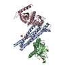
Refinement TLS group
ID
Refine-ID
Refine TLS-ID
Selection details
1
X-RAY DIFFRACTION
1
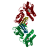
chain 'A'
2
X-RAY DIFFRACTION
2
chain 'B'
3
X-RAY DIFFRACTION
3
chain 'C'
4
X-RAY DIFFRACTION
4
chain 'D'
+
About Yorodumi
-
News
-
Feb 9, 2022. New format data for meta-information of EMDB entries
New format data for meta-information of EMDB entries
Version 3 of the EMDB header file is now the official format.
The previous official version 1.9 will be removed from the archive.
In the structure databanks used in Yorodumi, some data are registered as the other names, "COVID-19 virus" and "2019-nCoV". Here are the details of the virus and the list of structure data.
Jan 31, 2019. EMDB accession codes are about to change! (news from PDBe EMDB page)
EMDB accession codes are about to change! (news from PDBe EMDB page)
The allocation of 4 digits for EMDB accession codes will soon come to an end. Whilst these codes will remain in use, new EMDB accession codes will include an additional digit and will expand incrementally as the available range of codes is exhausted. The current 4-digit format prefixed with “EMD-” (i.e. EMD-XXXX) will advance to a 5-digit format (i.e. EMD-XXXXX), and so on. It is currently estimated that the 4-digit codes will be depleted around Spring 2019, at which point the 5-digit format will come into force.
The EM Navigator/Yorodumi systems omit the EMD- prefix.
Related info.:Q: What is EMD? / ID/Accession-code notation in Yorodumi/EM Navigator
Yorodumi is a browser for structure data from EMDB, PDB, SASBDB, etc.
This page is also the successor to EM Navigator detail page, and also detail information page/front-end page for Omokage search.
The word "yorodu" (or yorozu) is an old Japanese word meaning "ten thousand". "mi" (miru) is to see.
Related info.:EMDB / PDB / SASBDB / Comparison of 3 databanks / Yorodumi Search / Aug 31, 2016. New EM Navigator & Yorodumi / Yorodumi Papers / Jmol/JSmol / Function and homology information / Changes in new EM Navigator and Yorodumi
 Movie
Movie Controller


 Open data
 Basic information
 Components
 Keywords
 Function and homology information
 Homo sapiens (human)
 X-RAY DIFFRACTION /
  Authors
 Citation
 Structure visualization
 Downloads & links
 Other downloads
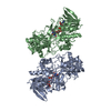


 PDBj
PDBj


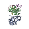
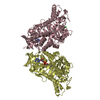
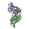
 Assembly
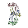


 Mass: 35.453 Da / Num. of mol.: 2 / Source method: obtained synthetically / Formula: Cl
 Mass: 18.015 Da / Num. of mol.: 120 / Source method: isolated from a natural source / Formula: H2O
 Sample preparation
 / Beamline: I911-3 / Wavelength: 1 Å
 Processing