 Movie
Movie Controller

[English] 日本語
 Yorodumi
- PDB-3u23: Atomic resolution crystal structure of the 2nd SH3 domain from hu... -
+ Open data

- Basic information
| Entry | Database: PDB / ID: 3u23 | ||||||
|---|---|---|---|---|---|---|---|
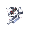
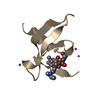
| Title | Atomic resolution crystal structure of the 2nd SH3 domain from human CD2AP (CMS) in complex with a proline-rich peptide from human RIN3 | ||||||
 Components Components |
| ||||||
 Keywords Keywords | PROTEIN BINDING / Structural Genomics / Structural Genomics Consortium / SGC / Beta-barrel / Adaptor protein | ||||||
| Function / homology |  Function and homology information Function and homology informationnegative regulation of mast cell chemotaxis / response to glial cell derived neurotrophic factor / negative regulation of small GTPase mediated signal transduction / transforming growth factor beta1 production / localization of cell / Rab protein signal transduction / negative regulation of transforming growth factor beta1 production / slit diaphragm / response to transforming growth factor beta / podocyte differentiation ...negative regulation of mast cell chemotaxis / response to glial cell derived neurotrophic factor / negative regulation of small GTPase mediated signal transduction / transforming growth factor beta1 production / localization of cell / Rab protein signal transduction / negative regulation of transforming growth factor beta1 production / slit diaphragm / response to transforming growth factor beta / podocyte differentiation / regulation of vesicle size / immunological synapse formation / endothelium development / nerve growth factor signaling pathway / cell-cell adhesion mediated by cadherin / collateral sprouting / protein heterooligomerization / renal albumin absorption / substrate-dependent cell migration, cell extension / phosphatidylinositol 3-kinase regulatory subunit binding / membrane organization / RAB GEFs exchange GTP for GDP on RABs / filopodium assembly / cell-cell junction organization / negative regulation of receptor internalization / podosome / Nephrin family interactions / clathrin binding / maintenance of blood-brain barrier / nuclear envelope lumen / glucose import / cell leading edge / filamentous actin / neurotrophin TRK receptor signaling pathway / endocytic vesicle / centriolar satellite / protein secretion / lymph node development / adipose tissue development / stress-activated MAPK cascade / ruffle / ERK1 and ERK2 cascade / actin filament polymerization / phosphatidylinositol 3-kinase/protein kinase B signal transduction / GTPase activator activity / guanyl-nucleotide exchange factor activity / trans-Golgi network membrane / liver development / actin filament organization / positive regulation of protein secretion / lipid metabolic process / regulation of actin cytoskeleton organization / synapse organization / response to insulin / response to virus / protein catabolic process / neuromuscular junction / regulation of synaptic plasticity / structural constituent of cytoskeleton / fibrillar center / small GTPase binding / response to wounding / positive regulation of protein localization to nucleus / SH3 domain binding / actin filament binding / endocytosis / male gonad development / actin cytoskeleton / cell migration / late endosome / T cell receptor signaling pathway / growth cone / cytoplasmic vesicle / protein-containing complex assembly / vesicle / negative regulation of neuron apoptotic process / response to oxidative stress / cell population proliferation / early endosome / cadherin binding / inflammatory response / cell division / axon / neuronal cell body / dendrite / apoptotic process / signal transduction / extracellular exosome / identical protein binding / plasma membrane / cytoplasm / cytosol Similarity search - Function | ||||||
| Biological species |  Homo sapiens (human) Homo sapiens (human) | ||||||
| Method |  X-RAY DIFFRACTION / SYNCHROTRON / MOLECULAR REPLACEMENT / Resolution: 1.11 Å X-RAY DIFFRACTION / SYNCHROTRON / MOLECULAR REPLACEMENT / Resolution: 1.11 Å | ||||||
 Authors Authors | Simister, P.C. / Rouka, E. / Janning, M. / Muniz, J.R.C. / Kirsch, K.H. / Knapp, S. / von Delft, F. / Filippakopoulos, P. / Arrowsmith, C.H. / Krojer, T. ...Simister, P.C. / Rouka, E. / Janning, M. / Muniz, J.R.C. / Kirsch, K.H. / Knapp, S. / von Delft, F. / Filippakopoulos, P. / Arrowsmith, C.H. / Krojer, T. / Edwards, A.M. / Weigelt, J. / Bountra, C. / Feller, S.M. / Structural Genomics Consortium (SGC) | ||||||
 Citation Citation | Journal: J.Biol.Chem. / Year: 2015 Title: Differential Recognition Preferences of the Three Src Homology 3 (SH3) Domains from the Adaptor CD2-associated Protein (CD2AP) and Direct Association with Ras and Rab Interactor 3 (RIN3). Authors: Rouka, E. / Simister, P.C. / Janning, M. / Kumbrink, J. / Konstantinou, T. / Muniz, J.R. / Joshi, D. / O'Reilly, N. / Volkmer, R. / Ritter, B. / Knapp, S. / von Delft, F. / Kirsch, K.H. / Feller, S.M. | ||||||
| History |
|
- Structure visualization
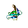
| Structure viewer | Molecule: MolmilJmol/JSmol |
|---|
- Downloads & links
-Download
| PDBx/mmCIF format | 3u23.cif.gz | 48.2 KB | Display | PDBx/mmCIF format |
|---|---|---|---|---|
| PDB format | pdb3u23.ent.gz | 33.7 KB | Display | PDB format |
| PDBx/mmJSON format | 3u23.json.gz | Tree view | PDBx/mmJSON format | |
| Others |  Other downloads Other downloads |
-Validation report
| Summary document | 3u23_validation.pdf.gz | 425.6 KB | Display | wwPDB validaton report |
|---|---|---|---|---|
| Full document | 3u23_full_validation.pdf.gz | 425.6 KB | Display | |
| Data in XML | 3u23_validation.xml.gz | 6.1 KB | Display | |
| Data in CIF | 3u23_validation.cif.gz | 7.7 KB | Display | |
| Arichive directory | https://data.pdbj.org/pub/pdb/validation_reports/u2/3u23ftp://data.pdbj.org/pub/pdb/validation_reports/u2/3u23 | HTTPS FTP |
-Related structure data
| Related structure data |  4wciC  1oebS 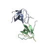 2ak5S 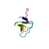 2feiS 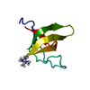 2g6fS 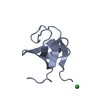 3iqlS S: Starting model for refinement C: citing same article ( |
|---|---|
| Similar structure data |


-Links
 PDBj
PDBj
- Assembly
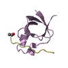
| Deposited unit | 
| ||||||||
|---|---|---|---|---|---|---|---|---|---|
| 1 |
| ||||||||
| Unit cell |
|
-Components
| #1: Protein | Mass: 7444.361 Da / Num. of mol.: 1 / Fragment: unp residues 109-168 Source method: isolated from a genetically manipulated source Source: (gene. exp.) Homo sapiens (human) / Gene: CD2AP / Production host:  |
|---|---|
| #2: Protein/peptide | Mass: 1803.201 Da / Num. of mol.: 1 / Fragment: unp residues 452-467 / Source method: obtained synthetically / Details: Synthesised peptide / Source: (synth.) Homo sapiens (human) / References: UniProt: Q8TB24 |
| #3: Chemical | ChemComp-EDO /   Mass: 62.068 Da / Num. of mol.: 1 / Source method: obtained synthetically / Formula: C2H6O2 Mass: 62.068 Da / Num. of mol.: 1 / Source method: obtained synthetically / Formula: C2H6O2 |
| #4: Water | ChemComp-HOH /  Mass: 18.015 Da / Num. of mol.: 86 / Source method: isolated from a natural source / Formula: H2O Mass: 18.015 Da / Num. of mol.: 86 / Source method: isolated from a natural source / Formula: H2O |
-Experimental details
-Experiment
| Experiment | Method: X-RAY DIFFRACTION / Number of used crystals: 1 |
|---|
- Sample preparation
| Crystal | Density Matthews: 1.8 Å3/Da / Density % sol: 31.7 % |
|---|---|
| Crystal grow | Temperature: 298 K / Method: vapor diffusion, sitting drop / pH: 7.5 Details: 0.1 M HEPES pH 7.5, 1.4 M tri-sodium citrate dihydrate, VAPOR DIFFUSION, SITTING DROP, temperature 298K |
-Data collection
| Diffraction | Mean temperature: 100 K |
|---|---|
| Diffraction source | Source: SYNCHROTRON / Site: Diamond  / Beamline: I03 / Wavelength: 0.97625 Å / Beamline: I03 / Wavelength: 0.97625 Å |
| Detector | Type: PSI PILATUS 6M / Detector: PIXEL / Date: Feb 13, 2011 |
| Radiation | Monochromator: Double crystal / Protocol: SINGLE WAVELENGTH / Monochromatic (M) / Laue (L): M / Scattering type: x-ray |
| Radiation wavelength | Wavelength: 0.97625 Å / Relative weight: 1 |
| Reflection | Resolution: 1.11→27.43 Å / Num. all: 26161 / Num. obs: 24275 / % possible obs: 92.8 % / Observed criterion σ(F): -3 / Observed criterion σ(I): -3 / Redundancy: 6 % / Biso Wilson estimate: 13.68 Å2 / Rmerge(I) obs: 0.056 / Net I/σ(I): 14.98 |
| Reflection shell | Resolution: 1.11→1.14 Å / Redundancy: 2.7 % / Rmerge(I) obs: 0.521 / Mean I/σ(I) obs: 2.24 / % possible all: 59.9 |
- Processing
| Software |
| ||||||||||||||||||||||||||||||||||||||||||||||||||||||||||||||||||||||||||||||||||||||||||||||||||||||||||||||||||||||||||||||||||||||||||||||||||||||||||||||||||||||||||
|---|---|---|---|---|---|---|---|---|---|---|---|---|---|---|---|---|---|---|---|---|---|---|---|---|---|---|---|---|---|---|---|---|---|---|---|---|---|---|---|---|---|---|---|---|---|---|---|---|---|---|---|---|---|---|---|---|---|---|---|---|---|---|---|---|---|---|---|---|---|---|---|---|---|---|---|---|---|---|---|---|---|---|---|---|---|---|---|---|---|---|---|---|---|---|---|---|---|---|---|---|---|---|---|---|---|---|---|---|---|---|---|---|---|---|---|---|---|---|---|---|---|---|---|---|---|---|---|---|---|---|---|---|---|---|---|---|---|---|---|---|---|---|---|---|---|---|---|---|---|---|---|---|---|---|---|---|---|---|---|---|---|---|---|---|---|---|---|---|---|---|---|
| Refinement | Method to determine structure: MOLECULAR REPLACEMENT Starting model: pdb entries 2G6F, 1OEB, 2AK5, 2FEI, 3IQL Resolution: 1.11→27.43 Å / Cor.coef. Fo:Fc: 0.981 / Cor.coef. Fo:Fc free: 0.978 / SU B: 1.332 / SU ML: 0.028 / Cross valid method: THROUGHOUT / ESU R: 0.035 / ESU R Free: 0.033 / Stereochemistry target values: MAXIMUM LIKELIHOOD / Details: HYDROGENS HAVE BEEN ADDED IN THE RIDING POSITIONS
| ||||||||||||||||||||||||||||||||||||||||||||||||||||||||||||||||||||||||||||||||||||||||||||||||||||||||||||||||||||||||||||||||||||||||||||||||||||||||||||||||||||||||||
| Solvent computation | Ion probe radii: 0.8 Å / Shrinkage radii: 0.8 Å / VDW probe radii: 1.4 Å / Solvent model: MASK | ||||||||||||||||||||||||||||||||||||||||||||||||||||||||||||||||||||||||||||||||||||||||||||||||||||||||||||||||||||||||||||||||||||||||||||||||||||||||||||||||||||||||||
| Displacement parameters | Biso mean: 15.91 Å2
| ||||||||||||||||||||||||||||||||||||||||||||||||||||||||||||||||||||||||||||||||||||||||||||||||||||||||||||||||||||||||||||||||||||||||||||||||||||||||||||||||||||||||||
| Refinement step | Cycle: LAST / Resolution: 1.11→27.43 Å
| ||||||||||||||||||||||||||||||||||||||||||||||||||||||||||||||||||||||||||||||||||||||||||||||||||||||||||||||||||||||||||||||||||||||||||||||||||||||||||||||||||||||||||
| Refine LS restraints |
| ||||||||||||||||||||||||||||||||||||||||||||||||||||||||||||||||||||||||||||||||||||||||||||||||||||||||||||||||||||||||||||||||||||||||||||||||||||||||||||||||||||||||||
| LS refinement shell | Resolution: 1.11→1.139 Å / Total num. of bins used: 20
| ||||||||||||||||||||||||||||||||||||||||||||||||||||||||||||||||||||||||||||||||||||||||||||||||||||||||||||||||||||||||||||||||||||||||||||||||||||||||||||||||||||||||||
| Refinement TLS params. | Method: refined / Origin x: 6.401 Å / Origin y: 5.769 Å / Origin z: 2.544 Å
|
