 Movie
Movie Controller

[English] 日本語
 Yorodumi
- PDB-6v2d: Crystal Structure of chromodomain of CDYL2 in complex with inhibi... -
+ Open data

- Basic information
| Entry | Database: PDB / ID: 6v2d | ||||||
|---|---|---|---|---|---|---|---|
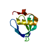
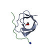
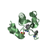
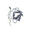
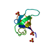
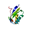
| Title | Crystal Structure of chromodomain of CDYL2 in complex with inhibitor UNC3866 | ||||||
 Components Components |
| ||||||
 Keywords Keywords | GENE REGULATION / structural genomics / Structural Genomics Consortium / SGC | ||||||
| Function / homology |  Function and homology information Function and homology informationhistone H3K27me3 reader activity / histone H3K9me2/3 reader activity / transcription corepressor activity / nucleus Similarity search - Function | ||||||
| Biological species |  Homo sapiens (human) Homo sapiens (human)synthetic construct (others) | ||||||
| Method |  X-RAY DIFFRACTION / MOLECULAR REPLACEMENT / Resolution: 2.1 Å X-RAY DIFFRACTION / MOLECULAR REPLACEMENT / Resolution: 2.1 Å | ||||||
 Authors Authors | Liu, Y. / Tempel, W. / Bountra, C. / Arrowsmith, C.H. / Edwards, A.M. / Min, J. / Structural Genomics Consortium (SGC) | ||||||
 Citation Citation | Journal: Cell Chem Biol / Year: 2020 Title: Structural Basis for the Binding Selectivity of Human CDY Chromodomains. Authors: Dong, C. / Liu, Y. / Lyu, T.J. / Beldar, S. / Lamb, K.N. / Tempel, W. / Li, Y. / Li, Z. / James, L.I. / Qin, S. / Wang, Y. / Min, J. | ||||||
| History |
|
- Structure visualization
| Structure viewer | Molecule: MolmilJmol/JSmol |
|---|
- Downloads & links
-Download
| PDBx/mmCIF format | 6v2d.cif.gz | 120.6 KB | Display | PDBx/mmCIF format |
|---|---|---|---|---|
| PDB format | pdb6v2d.ent.gz | 77.1 KB | Display | PDB format |
| PDBx/mmJSON format | 6v2d.json.gz | Tree view | PDBx/mmJSON format | |
| Others |  Other downloads Other downloads |
-Validation report
| Arichive directory | https://data.pdbj.org/pub/pdb/validation_reports/v2/6v2dftp://data.pdbj.org/pub/pdb/validation_reports/v2/6v2d | HTTPS FTP |
|---|
-Related structure data
| Related structure data |  2mj8C 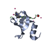 6v2hC  6v2rC  6v2sC 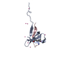 6v3nC 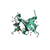 6v41C  6v8wC  5epjS 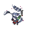 5epkS S: Starting model for refinement C: citing same article ( |
|---|---|
| Similar structure data | |
| Experimental dataset #1 | Data reference: 10.5281/zenodo.3532171 / Data set type: diffraction image data |


-Links
 PDBj
PDBj- Assembly
| Deposited unit | 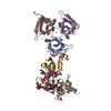
| ||||||||||
|---|---|---|---|---|---|---|---|---|---|---|---|
| 1 | 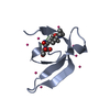
| ||||||||||
| 2 | 
| ||||||||||
| 3 | 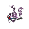
| ||||||||||
| 4 | 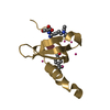
| ||||||||||
| 5 | 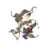
| ||||||||||
| 6 | 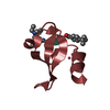
| ||||||||||
| Unit cell |
|
-Components
| #1: Protein | Mass: 7695.596 Da / Num. of mol.: 6 / Fragment: Chromodomain Source method: isolated from a genetically manipulated source Source: (gene. exp.) Homo sapiens (human) / Gene: CDYL2 / Plasmid: pET28-MHL / Production host:  #2: Protein/peptide |   Type: Oligopeptide / Class: Inhibitor / Mass: 795.020 Da / Num. of mol.: 6 / Source method: obtained synthetically / Source: (synth.) synthetic construct (others) / References: UNC3866 Type: Oligopeptide / Class: Inhibitor / Mass: 795.020 Da / Num. of mol.: 6 / Source method: obtained synthetically / Source: (synth.) synthetic construct (others) / References: UNC3866#3: Chemical | ChemComp-UNX /   Mass: 18.015 Da / Num. of mol.: 28 / Source method: isolated from a natural source Mass: 18.015 Da / Num. of mol.: 28 / Source method: isolated from a natural source#4: Water | ChemComp-HOH / |  Mass: 18.015 Da / Num. of mol.: 150 / Source method: isolated from a natural source / Formula: H2O Mass: 18.015 Da / Num. of mol.: 150 / Source method: isolated from a natural source / Formula: H2OHas ligand of interest | Y | |
|---|
-Experimental details
-Experiment
| Experiment | Method: X-RAY DIFFRACTION / Number of used crystals: 1 |
|---|
- Sample preparation
| Crystal | Density Matthews: 2.2 Å3/Da / Density % sol: 44.3 % |
|---|---|
| Crystal grow | Temperature: 293 K / Method: vapor diffusion, sitting drop / pH: 7.5 Details: 25% P3350, 0.2M ammonium acetate, 0.1M HEPES, pH 7.5 |
-Data collection
| Diffraction | Mean temperature: 100 K / Serial crystal experiment: N | ||||||||||||||||||||||||
|---|---|---|---|---|---|---|---|---|---|---|---|---|---|---|---|---|---|---|---|---|---|---|---|---|---|
| Diffraction source | Source: ROTATING ANODE / Type: RIGAKU FR-E / Wavelength: 1.5418 Å | ||||||||||||||||||||||||
| Detector | Type: RIGAKU SATURN A200 / Detector: CCD / Date: Apr 23, 2014 | ||||||||||||||||||||||||
| Radiation | Protocol: SINGLE WAVELENGTH / Monochromatic (M) / Laue (L): M / Scattering type: x-ray | ||||||||||||||||||||||||
| Radiation wavelength | Wavelength: 1.5418 Å / Relative weight: 1 | ||||||||||||||||||||||||
| Reflection | Resolution: 2→38.43 Å / Num. obs: 28615 / % possible obs: 92.1 % / Redundancy: 6.9 % / Biso Wilson estimate: 25.81 Å2 / CC1/2: 0.998 / Rmerge(I) obs: 0.108 / Rrim(I) all: 0.116 / Net I/σ(I): 14.7 | ||||||||||||||||||||||||
| Reflection shell |
|
- Processing
| Software |
| ||||||||||||||||||||||||||||||||||||||||||||||||||||||||||||||||||||||
|---|---|---|---|---|---|---|---|---|---|---|---|---|---|---|---|---|---|---|---|---|---|---|---|---|---|---|---|---|---|---|---|---|---|---|---|---|---|---|---|---|---|---|---|---|---|---|---|---|---|---|---|---|---|---|---|---|---|---|---|---|---|---|---|---|---|---|---|---|---|---|---|
| Refinement | Method to determine structure: MOLECULAR REPLACEMENT Starting model: preliminary coordinates of PDB entries 5EPJ and 5EPK Resolution: 2.1→38.05 Å / SU ML: 0.259 / Cross valid method: FREE R-VALUE / σ(F): 1.25 / Phase error: 27.1204 Stereochemistry target values: GeoStd + Monomer Library + CDL v1.2
| ||||||||||||||||||||||||||||||||||||||||||||||||||||||||||||||||||||||
| Solvent computation | Shrinkage radii: 0.9 Å / VDW probe radii: 1.11 Å / Solvent model: FLAT BULK SOLVENT MODEL | ||||||||||||||||||||||||||||||||||||||||||||||||||||||||||||||||||||||
| Displacement parameters | Biso mean: 25.65 Å2 | ||||||||||||||||||||||||||||||||||||||||||||||||||||||||||||||||||||||
| Refinement step | Cycle: LAST / Resolution: 2.1→38.05 Å
| ||||||||||||||||||||||||||||||||||||||||||||||||||||||||||||||||||||||
| Refine LS restraints |
| ||||||||||||||||||||||||||||||||||||||||||||||||||||||||||||||||||||||
| LS refinement shell |
|
