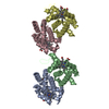
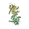
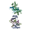
THIS ENTRY 3P5J REFLECTS AN ALTERNATIVE MODELING OF THE ORIGINAL STRUCTURAL DATA R3KIOSF DETERMINED ...THIS ENTRY 3P5J REFLECTS AN ALTERNATIVE MODELING OF THE ORIGINAL STRUCTURAL DATA R3KIOSF DETERMINED BY AUTHORS OF THE PDB ENTRY 3KIO: N.Shaban,D.Harvey,F.W.Perrino,T.Hollis.
Method to determine structure: MAD / Resolution: 2.9→24.61 Å / Cor.coef. Fo:Fc: 0.8969 / Cor.coef. Fo:Fc free: 0.8348 / Cross valid method: THROUGHOUT / σ(F): 0 / Stereochemistry target values: Engh & Huber Details: This is a re-refinement of the murine RNase H2 coordinates previously deposited by Shaban et al., 2010 (PDB ID 3KIO)
Rfactor
Num. reflection
% reflection
Selection details
Rfree
0.2716
925
5.2 %
RANDOM
Rwork
0.21
-
-
-
obs
0.2132
17777
-
-
Displacement parameters
Biso mean: 62.48 Å2
Baniso -1
Baniso -2
Baniso -3
1-
13.0671 Å2
0 Å2
0 Å2
2-
-
-4.4747 Å2
0 Å2
3-
-
-
-8.5923 Å2
Refine analyze
Luzzati coordinate error obs: 0.405 Å
Refinement step
Cycle: LAST / Resolution: 2.9→24.61 Å
Protein
Nucleic acid
Ligand
Solvent
Total
Num. atoms
4016
0
0
0
4016
Refine LS restraints
Refine-ID
Type
Dev ideal
Number
Restraint function
Weight
X-RAY DIFFRACTION
t_bond_d
0.011
4116
HARMONIC
2
X-RAY DIFFRACTION
t_angle_deg
1.28
5614
HARMONIC
2
X-RAY DIFFRACTION
t_dihedral_angle_d
1269
SINUSOIDAL
2
X-RAY DIFFRACTION
t_incorr_chiral_ct
X-RAY DIFFRACTION
t_pseud_angle
X-RAY DIFFRACTION
t_trig_c_planes
68
HARMONIC
2
X-RAY DIFFRACTION
t_gen_planes
630
HARMONIC
5
X-RAY DIFFRACTION
t_it
4116
HARMONIC
20
X-RAY DIFFRACTION
t_nbd
X-RAY DIFFRACTION
t_omega_torsion
3.09
X-RAY DIFFRACTION
t_other_torsion
21.53
X-RAY DIFFRACTION
t_improper_torsion
X-RAY DIFFRACTION
t_chiral_improper_torsion
548
5
X-RAY DIFFRACTION
t_sum_occupancies
X-RAY DIFFRACTION
t_utility_distance
X-RAY DIFFRACTION
t_utility_angle
X-RAY DIFFRACTION
t_utility_torsion
X-RAY DIFFRACTION
t_ideal_dist_contact
4644
4
LS refinement shell
Resolution: 2.9→3.08 Å / Total num. of bins used: 9
Rfactor
Num. reflection
% reflection
Rfree
0.2934
163
5.8 %
Rwork
0.229
2649
-
all
0.2326
2812
-
Refinement TLS params.
Method: refined / Refine-ID: X-RAY DIFFRACTION
ID
L11 (°2)
L12 (°2)
L13 (°2)
L22 (°2)
L23 (°2)
L33 (°2)
S11 (Å °)
S12 (Å °)
S13 (Å °)
S21 (Å °)
S22 (Å °)
S23 (Å °)
S31 (Å °)
S32 (Å °)
S33 (Å °)
T11 (Å2)
T12 (Å2)
T13 (Å2)
T22 (Å2)
T23 (Å2)
T33 (Å2)
Origin x (Å)
Origin y (Å)
Origin z (Å)
1
1.1145
-0.2948
0.7102
1.2523
-0.5222
2.3424
-0.0432
-0.0372
-0.1281
0.0149
0.0176
-0.0206
-0.0651
0.1222
0.0257
0.1858
-0.0251
-0.0022
-0.2376
-0.0287
-0.304
73.2007
9.6824
22.1632
2
5.9326
2.5634
-0.1113
0.0013
-0.3888
0.4014
-0.023
0.1306
-0.1646
-0.1336
0.0269
0.5442
0.0586
-0.3411
-0.004
-0.304
-0.0503
-0.042
-0.2907
-0.0841
0.304
22.3709
-7.1893
3.4824
3
4.5519
0.2111
0.9306
3.6213
0.8695
1.5601
0.1005
0.2979
-0.4617
-0.1266
-0.0983
0.5442
0.197
-0.1822
-0.0021
0.0704
-0.0255
-0.0155
-0.2576
-0.0167
-0.1683
45.4622
-2.7782
4.2073
Refinement TLS group
ID
Refine-ID
Refine TLS-ID
Selection details
Auth asym-ID
Auth seq-ID
1
X-RAY DIFFRACTION
1
{ A|* }
A
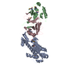
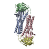
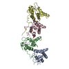
10 - 299
2
X-RAY DIFFRACTION
2
{ B|* }
B
11 - 231
3
X-RAY DIFFRACTION
3
{ C|* }
C
14 - 162
+
About Yorodumi
-
News
-
Feb 9, 2022. New format data for meta-information of EMDB entries
New format data for meta-information of EMDB entries
Version 3 of the EMDB header file is now the official format.
The previous official version 1.9 will be removed from the archive.
In the structure databanks used in Yorodumi, some data are registered as the other names, "COVID-19 virus" and "2019-nCoV". Here are the details of the virus and the list of structure data.
Jan 31, 2019. EMDB accession codes are about to change! (news from PDBe EMDB page)
EMDB accession codes are about to change! (news from PDBe EMDB page)
The allocation of 4 digits for EMDB accession codes will soon come to an end. Whilst these codes will remain in use, new EMDB accession codes will include an additional digit and will expand incrementally as the available range of codes is exhausted. The current 4-digit format prefixed with “EMD-” (i.e. EMD-XXXX) will advance to a 5-digit format (i.e. EMD-XXXXX), and so on. It is currently estimated that the 4-digit codes will be depleted around Spring 2019, at which point the 5-digit format will come into force.
The EM Navigator/Yorodumi systems omit the EMD- prefix.
Related info.:Q: What is EMD? / ID/Accession-code notation in Yorodumi/EM Navigator
Yorodumi is a browser for structure data from EMDB, PDB, SASBDB, etc.
This page is also the successor to EM Navigator detail page, and also detail information page/front-end page for Omokage search.
The word "yorodu" (or yorozu) is an old Japanese word meaning "ten thousand". "mi" (miru) is to see.
Related info.:EMDB / PDB / SASBDB / Comparison of 3 databanks / Yorodumi Search / Aug 31, 2016. New EM Navigator & Yorodumi / Yorodumi Papers / Jmol/JSmol / Function and homology information / Changes in new EM Navigator and Yorodumi
 Movie
Movie Controller

 Yorodumi
 Open data
 Basic information
 Components
 Keywords
 Function and homology information
 X-RAY DIFFRACTION /
  Authors
 Citation
 Structure visualization
 Downloads & links
 Other downloads
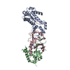


 PDBj
PDBj
 Assembly

 Sample preparation
 Processing