 Movie
Movie Controller

[English] 日本語
 Yorodumi
- PDB-3lh2: Crystal structure of HIV epitope-scaffold 4E10_1VI7A_S0_002_N 4E1... -
+ Open data

- Basic information
| Entry | Database: PDB / ID: 3lh2 | ||||||
|---|---|---|---|---|---|---|---|
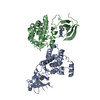
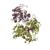
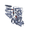
| Title | Crystal structure of HIV epitope-scaffold 4E10_1VI7A_S0_002_N 4E10 Fv complex | ||||||
 Components Components |
| ||||||
 Keywords Keywords |  IMMUNE SYSTEM / EPITOPE-SCAFFOLD IMMUNE SYSTEM / EPITOPE-SCAFFOLD | ||||||
| Function / homology | Alpha-Beta Plaits - #240 / Alpha-Beta Plaits / Immunoglobulins / Immunoglobulin-like / Sandwich / 2-Layer Sandwich / Mainly Beta / Alpha Beta Function and homology information Function and homology information | ||||||
| Biological species | ARTIFICIAL GENE (others) Homo sapiens (human) Homo sapiens (human) | ||||||
| Method | X-RAY DIFFRACTION / MOLECULAR REPLACEMENT / Resolution: 2.65 Å | ||||||
 Authors Authors | Holmes, M.A. | ||||||
 Citation Citation | Journal: Structure / Year: 2010 Title: Computational Design of Epitope-Scaffolds Allows Induction of Antibodies Specific for a Poorly Immunogenic HIV Vaccine Epitope. Authors: Correia, B.E. / Ban, Y.E. / Holmes, M.A. / Xu, H. / Ellingson, K. / Kraft, Z. / Carrico, C. / Boni, E. / Sather, D.N. / Zenobia, C. / Burke, K.Y. / Bradley-Hewitt, T. / Bruhn-Johannsen, J.F. ...Authors: Correia, B.E. / Ban, Y.E. / Holmes, M.A. / Xu, H. / Ellingson, K. / Kraft, Z. / Carrico, C. / Boni, E. / Sather, D.N. / Zenobia, C. / Burke, K.Y. / Bradley-Hewitt, T. / Bruhn-Johannsen, J.F. / Kalyuzhniy, O. / Baker, D. / Strong, R.K. / Stamatatos, L. / Schief, W.R. | ||||||
| History |
|
- Structure visualization
| Structure viewer | Molecule: MolmilJmol/JSmol |
|---|
- Downloads & links
-Download
| PDBx/mmCIF format | 3lh2.cif.gz | 231.6 KB | Display | PDBx/mmCIF format |
|---|---|---|---|---|
| PDB format | pdb3lh2.ent.gz | 186.3 KB | Display | PDB format |
| PDBx/mmJSON format | 3lh2.json.gz | Tree view | PDBx/mmJSON format | |
| Others |  Other downloads Other downloads |
-Validation report
| Arichive directory | https://data.pdbj.org/pub/pdb/validation_reports/lh/3lh2ftp://data.pdbj.org/pub/pdb/validation_reports/lh/3lh2 | HTTPS FTP |
|---|
-Related structure data
| Related structure data | 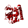 3lefC 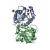 3lf6C 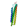 3lf9C 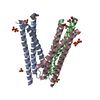 3lg7C 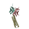 3lhpC 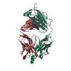 1tzgS C: citing same article ( S: Starting model for refinement |
|---|---|
| Similar structure data |


-Links
 PDBj
PDBj

- Assembly
| Deposited unit | 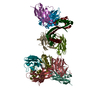
| ||||||||
|---|---|---|---|---|---|---|---|---|---|
| 1 | 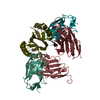
| ||||||||
| 2 | 
| ||||||||
| Unit cell |
|
-Components
| #1: Protein | Mass: 8455.588 Da / Num. of mol.: 4 Source method: isolated from a genetically manipulated source Details: The author states that THE EPITOPE-SCAFFOLD IS BASED on the ribosome recycling factor from Vibrio parahaemolyticus (PDB ID 1IS1). Source: (gene. exp.) ARTIFICIAL GENE (others) / Plasmid: PET29 / Production host:  Escherichia coli (E. coli) / Strain (production host): BL21(DE3) STAR Escherichia coli (E. coli) / Strain (production host): BL21(DE3) STAR#2: Antibody | Mass: 14712.454 Da / Num. of mol.: 4 / Mutation: W104A Source method: isolated from a genetically manipulated source Source: (gene. exp.) Homo sapiens (human) / Plasmid: PET22B / Production host: Escherichia coli (E. coli) / Strain (production host): BL21(DE3) RIL#3: Antibody | Mass: 12304.698 Da / Num. of mol.: 4 Source method: isolated from a genetically manipulated source Source: (gene. exp.) Homo sapiens (human) / Plasmid: PET22B / Production host: Escherichia coli (E. coli) / Strain (production host): BL21(DE3) RIL#4: Water | ChemComp-HOH / | Water Mass: 18.015 Da / Num. of mol.: 132 / Source method: isolated from a natural source / Formula: H2O Mass: 18.015 Da / Num. of mol.: 132 / Source method: isolated from a natural source / Formula: H2O |
|---|
-Experimental details
-Experiment
| Experiment | Method: X-RAY DIFFRACTION / Number of used crystals: 1 |
|---|
- Sample preparation
| Crystal | Density Matthews: 3.06 Å3/Da / Density % sol: 59.82 % |
|---|---|
| Crystal grow | Temperature: 298 K / Method: vapor diffusion, hanging drop / pH: 8 Details: Na acetate, imidazole, pH 8, VAPOR DIFFUSION, HANGING DROP, temperature 298K |
-Data collection
| Diffraction | Mean temperature: 107 K |
|---|---|
| Diffraction source | Source: ROTATING ANODE / Type: RIGAKU MICROMAX-007 / Wavelength: 1.54 Å |
| Detector | Type: RIGAKU SATURN 944+ / Detector: CCD / Date: Oct 22, 2008 / Details: Rigaku Varimax HF |
| Radiation | Monochromator: Mirrors / Protocol: SINGLE WAVELENGTH / Monochromatic (M) / Laue (L): M / Scattering type: x-ray |
| Radiation wavelength | Wavelength: 1.54 Å / Relative weight: 1 |
| Reflection | Resolution: 2.65→28.01 Å / Num. all: 49017 / Num. obs: 49017 / % possible obs: 98.9 % / Redundancy: 3.88 % / Biso Wilson estimate: 62.3 Å2 / Rmerge(I) obs: 0.095 / Net I/σ(I): 9.4 |
| Reflection shell | Resolution: 2.65→2.74 Å / Redundancy: 3.68 % / Rmerge(I) obs: 0.376 / Mean I/σ(I) obs: 2.7 / Num. unique all: 4883 / % possible all: 98.6 |
- Processing
| Software |
| ||||||||||||||||||||||||||||||||||||||||||||||||||||||||||||||||||||||||||||||||||||||||||||||||||||||||||||||||||||||||||||||||||||||||||||||||||||||||||||||||||||||||||
|---|---|---|---|---|---|---|---|---|---|---|---|---|---|---|---|---|---|---|---|---|---|---|---|---|---|---|---|---|---|---|---|---|---|---|---|---|---|---|---|---|---|---|---|---|---|---|---|---|---|---|---|---|---|---|---|---|---|---|---|---|---|---|---|---|---|---|---|---|---|---|---|---|---|---|---|---|---|---|---|---|---|---|---|---|---|---|---|---|---|---|---|---|---|---|---|---|---|---|---|---|---|---|---|---|---|---|---|---|---|---|---|---|---|---|---|---|---|---|---|---|---|---|---|---|---|---|---|---|---|---|---|---|---|---|---|---|---|---|---|---|---|---|---|---|---|---|---|---|---|---|---|---|---|---|---|---|---|---|---|---|---|---|---|---|---|---|---|---|---|---|---|
| Refinement | Method to determine structure: MOLECULAR REPLACEMENT Starting model: Computationally-derived model of the epitope-scaffold Fv complex, with the Fv based on PDB ID 1TZG. Resolution: 2.65→26.7 Å / Cor.coef. Fo:Fc: 0.921 / Cor.coef. Fo:Fc free: 0.878 / SU B: 11.214 / SU ML: 0.24 / Isotropic thermal model: Isotropic / Cross valid method: THROUGHOUT / ESU R: 0.578 / ESU R Free: 0.325 / Stereochemistry target values: Engh & Huber / Details: HYDROGENS HAVE BEEN ADDED IN THE RIDING POSITIONS
| ||||||||||||||||||||||||||||||||||||||||||||||||||||||||||||||||||||||||||||||||||||||||||||||||||||||||||||||||||||||||||||||||||||||||||||||||||||||||||||||||||||||||||
| Solvent computation | Ion probe radii: 0.8 Å / Shrinkage radii: 0.8 Å / VDW probe radii: 1.4 Å / Solvent model: BABINET MODEL WITH MASK | ||||||||||||||||||||||||||||||||||||||||||||||||||||||||||||||||||||||||||||||||||||||||||||||||||||||||||||||||||||||||||||||||||||||||||||||||||||||||||||||||||||||||||
| Displacement parameters | Biso mean: 47.111 Å2
| ||||||||||||||||||||||||||||||||||||||||||||||||||||||||||||||||||||||||||||||||||||||||||||||||||||||||||||||||||||||||||||||||||||||||||||||||||||||||||||||||||||||||||
| Refinement step | Cycle: LAST / Resolution: 2.65→26.7 Å
| ||||||||||||||||||||||||||||||||||||||||||||||||||||||||||||||||||||||||||||||||||||||||||||||||||||||||||||||||||||||||||||||||||||||||||||||||||||||||||||||||||||||||||
| Refine LS restraints |
| ||||||||||||||||||||||||||||||||||||||||||||||||||||||||||||||||||||||||||||||||||||||||||||||||||||||||||||||||||||||||||||||||||||||||||||||||||||||||||||||||||||||||||
| LS refinement shell | Resolution: 2.647→2.715 Å / Total num. of bins used: 20
|
