 Movie
Movie Controller


+ Open data

- Basic information
| Entry | Database: PDB / ID: 2ydv | ||||||
|---|---|---|---|---|---|---|---|
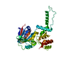
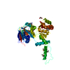
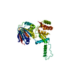
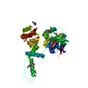
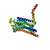
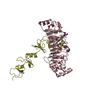
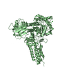
| Title | Thermostabilised HUMAN A2a Receptor with NECA bound | ||||||
 Components Components | ADENOSINE RECEPTOR A2A Adenosine A2A receptor Adenosine A2A receptor | ||||||
 Keywords Keywords | RECEPTOR / G PROTEIN COUPLED RECEPTOR / SEVEN-HELIX RECEPTOR / AGONIST BOUND FORM / THERMOSTABILISING POINT MUTATIONS / GPCR / 7TM RECEPTOR | ||||||
| Function / homology |  Function and homology information Function and homology informationpositive regulation of acetylcholine secretion, neurotransmission / positive regulation of circadian sleep/wake cycle, sleep / regulation of norepinephrine secretion / negative regulation of alpha-beta T cell activation / Adenosine P1 receptors / G protein-coupled adenosine receptor activity / G protein-coupled adenosine receptor signaling pathway / response to purine-containing compound / sensory perception / positive regulation of urine volume ...positive regulation of acetylcholine secretion, neurotransmission / positive regulation of circadian sleep/wake cycle, sleep / regulation of norepinephrine secretion / negative regulation of alpha-beta T cell activation / Adenosine P1 receptors / G protein-coupled adenosine receptor activity / G protein-coupled adenosine receptor signaling pathway / response to purine-containing compound / sensory perception / positive regulation of urine volume / NGF-independant TRKA activation / Surfactant metabolism / protein kinase C-activating G protein-coupled receptor signaling pathway / synaptic transmission, dopaminergic / synaptic transmission, cholinergic / inhibitory postsynaptic potential / negative regulation of vascular permeability / type 5 metabotropic glutamate receptor binding / positive regulation of glutamate secretion / blood circulation / intermediate filament / response to caffeine / eating behavior / presynaptic active zone / alpha-actinin binding / membrane depolarization / regulation of calcium ion transport / asymmetric synapse / axolemma / response to inorganic substance / cellular defense response / prepulse inhibition / phagocytosis / positive regulation of apoptotic signaling pathway / response to amphetamine / excitatory postsynaptic potential / presynaptic modulation of chemical synaptic transmission / positive regulation of synaptic transmission, glutamatergic / neuron projection morphogenesis / locomotory behavior / regulation of mitochondrial membrane potential / synaptic transmission, glutamatergic / central nervous system development / positive regulation of long-term synaptic potentiation / astrocyte activation / apoptotic signaling pathway / positive regulation of protein secretion / positive regulation of synaptic transmission, GABAergic / adenylate cyclase-modulating G protein-coupled receptor signaling pathway / adenylate cyclase-activating G protein-coupled receptor signaling pathway / negative regulation of inflammatory response / vasodilation / blood coagulation / cell-cell signaling / presynaptic membrane / G alpha (s) signalling events / postsynaptic membrane / negative regulation of neuron apoptotic process / calmodulin binding / response to xenobiotic stimulus / inflammatory response / negative regulation of cell population proliferation / dendrite / neuronal cell body / glutamatergic synapse / lipid binding / apoptotic process / protein-containing complex binding / regulation of DNA-templated transcription / enzyme binding / membrane / identical protein binding / plasma membraneSimilarity search - Function | ||||||
| Biological species |  HOMO SAPIENS (human) HOMO SAPIENS (human) | ||||||
| Method | X-RAY DIFFRACTION / SYNCHROTRON / MOLECULAR REPLACEMENT / Resolution: 2.6 Å | ||||||
 Authors Authors | Lebon, G. / Warne, T. / Edwards, P.C. / Bennett, K. / Langmead, C.J. / Leslie, A.G.W. / Tate, C.G. | ||||||
 Citation Citation | Journal: Nature / Year: 2011 Title: Agonist-Bound Adenosine A(2A) Receptor Structures Reveal Common Features of Gpcr Activation. Authors: Lebon, G. / Warne, T. / Edwards, P.C. / Bennett, K. / Langmead, C.J. / Leslie, A.G.W. / Tate, C.G. #1: Journal: J.Mol.Biol. / Year: 2011 Title: Thermostabilisation of an Agonist-Bound Conformation of the Human Adenosine A(2A) Receptor. Authors: Lebon, G. / Bennett, K. / Jazayeri, A. / Tate, C.G. | ||||||
| History |
|
- Structure visualization
| Structure viewer | Molecule: MolmilJmol/JSmol |
|---|
- Downloads & links
-Download
| PDBx/mmCIF format | 2ydv.cif.gz | 79.5 KB | Display | PDBx/mmCIF format |
|---|---|---|---|---|
| PDB format | pdb2ydv.ent.gz | 59 KB | Display | PDB format |
| PDBx/mmJSON format | 2ydv.json.gz | Tree view | PDBx/mmJSON format | |
| Others |  Other downloads Other downloads |
-Validation report
| Arichive directory | https://data.pdbj.org/pub/pdb/validation_reports/yd/2ydvftp://data.pdbj.org/pub/pdb/validation_reports/yd/2ydv | HTTPS FTP |
|---|
-Related structure data
| Related structure data |  2ydoC 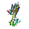 3emlS C: citing same article ( S: Starting model for refinement |
|---|---|
| Similar structure data |


-Links
 PDBj
PDBj


- Assembly
| Deposited unit | 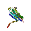
| ||||||||
|---|---|---|---|---|---|---|---|---|---|
| 1 |
| ||||||||
| Unit cell |
|
-Components
| #1: Protein | Adenosine A2A receptor / THERMOSTABILISED HUMAN A2A RECEPTOR Mass: 36022.773 Da / Num. of mol.: 1 / Fragment: RESIDUES 1-317 / Mutation: YES Source method: isolated from a genetically manipulated source Source: (gene. exp.) HOMO SAPIENS (human) / Tissue: BRAIN / Plasmid: PBACPAK8 / Cell line (production host): High Five / Production host:  TRICHOPLUSIA NI (cabbage looper) / References: UniProt: P29274 TRICHOPLUSIA NI (cabbage looper) / References: UniProt: P29274 | ||||||
|---|---|---|---|---|---|---|---|
| #2: Chemical | ChemComp-NEC /   Mass: 308.293 Da / Num. of mol.: 1 / Source method: obtained synthetically / Formula: C12H16N6O4 Mass: 308.293 Da / Num. of mol.: 1 / Source method: obtained synthetically / Formula: C12H16N6O4 | ||||||
| #3: Sugar | ChemComp-SOG / N-Octyl beta-D-thioglucopyranoside  Type: D-saccharide / Mass: 308.434 Da / Num. of mol.: 5 Type: D-saccharide / Mass: 308.434 Da / Num. of mol.: 5Source method: isolated from a genetically manipulated source Formula: C14H28O5S / Comment: detergent*YM #4: Water | ChemComp-HOH / | Water Mass: 18.015 Da / Num. of mol.: 27 / Source method: isolated from a natural source / Formula: H2O Mass: 18.015 Da / Num. of mol.: 27 / Source method: isolated from a natural source / Formula: H2OCompound details | ENGINEERED RESIDUE IN CHAIN A, LEU 48 TO ALA ENGINEERED RESIDUE IN CHAIN A, ALA 54 TO LEU ...ENGINEERED | Sequence details | THE CONSTRUCT WAS TRUNCATED AFTER RESIDUE 316 OF THE A2A SEQUENCE. CONSTRUCT CRYSTALLISED CONTAINS ...THE CONSTRUCT WAS TRUNCATED AFTER RESIDUE 316 OF THE A2A SEQUENCE. CONSTRUCT CRYSTALLIS | |
-Experimental details
-Experiment
| Experiment | Method: X-RAY DIFFRACTION / Number of used crystals: 1 |
|---|
- Sample preparation
| Crystal | Density Matthews: 4.01 Å3/Da / Density % sol: 69.3 % / Description: NONE |
|---|---|
| Crystal grow | pH: 6.4 Details: 0.05 M ADA NAOH, PH 6.4, 23.6% V/V PEG 400, 4% V/V 2-PROPANOL. |
-Data collection
| Diffraction | Mean temperature: 100 K |
|---|---|
| Diffraction source | Source: SYNCHROTRON / Site: ESRF  / Beamline: ID23-2 / Wavelength: 0.8726 / Beamline: ID23-2 / Wavelength: 0.8726 |
| Detector | Type: MARMOSAIC 225 mm CCD / Detector: CCD / Date: Nov 14, 2010 |
| Radiation | Protocol: SINGLE WAVELENGTH / Monochromatic (M) / Laue (L): M / Scattering type: x-ray |
| Radiation wavelength | Wavelength: 0.8726 Å / Relative weight: 1 |
| Reflection | Resolution: 2.6→49.41 Å / Num. obs: 17471 / % possible obs: 99.7 % / Observed criterion σ(I): 0 / Redundancy: 3.4 % / Biso Wilson estimate: 70.5 Å2 / Rmerge(I) obs: 0.08 / Net I/σ(I): 8.5 |
| Reflection shell | Resolution: 2.6→2.74 Å / Redundancy: 2.8 % / Rmerge(I) obs: 0.57 / Mean I/σ(I) obs: 1.7 / % possible all: 99.2 |
- Processing
| Software |
| ||||||||||||||||||||||||||||||||||||||||||||||||||||||||||||||||||||||||||||||||||||||||||||||||||||||||||||||||||||||||||||||||||||||||||||||||||||||||||||||||||||||||||||||||||||||
|---|---|---|---|---|---|---|---|---|---|---|---|---|---|---|---|---|---|---|---|---|---|---|---|---|---|---|---|---|---|---|---|---|---|---|---|---|---|---|---|---|---|---|---|---|---|---|---|---|---|---|---|---|---|---|---|---|---|---|---|---|---|---|---|---|---|---|---|---|---|---|---|---|---|---|---|---|---|---|---|---|---|---|---|---|---|---|---|---|---|---|---|---|---|---|---|---|---|---|---|---|---|---|---|---|---|---|---|---|---|---|---|---|---|---|---|---|---|---|---|---|---|---|---|---|---|---|---|---|---|---|---|---|---|---|---|---|---|---|---|---|---|---|---|---|---|---|---|---|---|---|---|---|---|---|---|---|---|---|---|---|---|---|---|---|---|---|---|---|---|---|---|---|---|---|---|---|---|---|---|---|---|---|---|
| Refinement | Method to determine structure: MOLECULAR REPLACEMENT Starting model: PDB ENTRY 3EML Resolution: 2.6→79.52 Å / Cor.coef. Fo:Fc: 0.938 / Cor.coef. Fo:Fc free: 0.929 / SU B: 9.904 / SU ML: 0.215 / Cross valid method: THROUGHOUT / ESU R: 0.374 / ESU R Free: 0.264 / Stereochemistry target values: MAXIMUM LIKELIHOOD Details: HYDROGENS HAVE BEEN ADDED IN THE RIDING POSITIONS. HYDROGENS HAVE BEEN USED IF PRESENT IN THE INPUT. U VALUES REFINED INDIVIDUALLY.
| ||||||||||||||||||||||||||||||||||||||||||||||||||||||||||||||||||||||||||||||||||||||||||||||||||||||||||||||||||||||||||||||||||||||||||||||||||||||||||||||||||||||||||||||||||||||
| Solvent computation | Ion probe radii: 0.8 Å / Shrinkage radii: 0.8 Å / VDW probe radii: 1.2 Å / Solvent model: MASK | ||||||||||||||||||||||||||||||||||||||||||||||||||||||||||||||||||||||||||||||||||||||||||||||||||||||||||||||||||||||||||||||||||||||||||||||||||||||||||||||||||||||||||||||||||||||
| Displacement parameters | Biso mean: 75.146 Å2
| ||||||||||||||||||||||||||||||||||||||||||||||||||||||||||||||||||||||||||||||||||||||||||||||||||||||||||||||||||||||||||||||||||||||||||||||||||||||||||||||||||||||||||||||||||||||
| Refinement step | Cycle: LAST / Resolution: 2.6→79.52 Å
| ||||||||||||||||||||||||||||||||||||||||||||||||||||||||||||||||||||||||||||||||||||||||||||||||||||||||||||||||||||||||||||||||||||||||||||||||||||||||||||||||||||||||||||||||||||||
| Refine LS restraints |
|