 Movie
Movie Controller

[English] 日本語
 Yorodumi
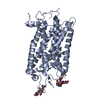
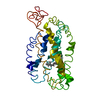
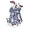
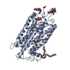
- PDB-2j4y: Crystal structure of a rhodopsin stabilizing mutant expressed in ... -
+ Open data

- Basic information
| Entry | Database: PDB / ID: 2j4y | ||||||
|---|---|---|---|---|---|---|---|
| Title | Crystal structure of a rhodopsin stabilizing mutant expressed in mammalian cells | ||||||
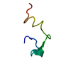
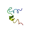
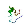
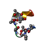
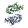
 Components Components | RHODOPSIN | ||||||
 Keywords Keywords | SIGNALING PROTEIN / CHROMOPHORE / LIPOPROTEIN / GLYCOPROTEIN / SENSORY TRANSDUCTION / PHOTORECEPTOR PROTEIN / INTEGRAL MEMBRANE PROTEIN / G-PROTEIN COUPLED RECEPTOR / VISION / MEMBRANE / RECEPTOR / PALMITATE / TRANSDUCER / RETINAL PROTEIN / PHOSPHORYLATION / PHOTORECEPTOR / TRANSMEMBRANE / VISUAL PIGMENT | ||||||
| Function / homology |  Function and homology informationOpsins / VxPx cargo-targeting to cilium / rod photoreceptor outer segment / rod bipolar cell differentiation / sperm head plasma membrane / podosome assembly / absorption of visible light / opsin binding / The canonical retinoid cycle in rods (twilight vision) / : ...Opsins / VxPx cargo-targeting to cilium / rod photoreceptor outer segment / rod bipolar cell differentiation / sperm head plasma membrane / podosome assembly / absorption of visible light / opsin binding / The canonical retinoid cycle in rods (twilight vision) / : / G protein-coupled photoreceptor activity / photoreceptor inner segment membrane / rhodopsin mediated signaling pathway / 11-cis retinal binding / cellular response to light stimulus / G protein-coupled receptor complex / Inactivation, recovery and regulation of the phototransduction cascade / phototransduction, visible light / thermotaxis / Activation of the phototransduction cascade / detection of temperature stimulus involved in thermoception / outer membrane / arrestin family protein binding / photoreceptor cell maintenance / photoreceptor outer segment membrane / G alpha (i) signalling events / response to light stimulus / phototransduction / photoreceptor outer segment / G-protein alpha-subunit binding / sperm midpiece / visual perception / guanyl-nucleotide exchange factor activity / microtubule cytoskeleton organization / photoreceptor disc membrane / cell-cell junction / gene expression / G protein-coupled receptor signaling pathway / Golgi membrane / zinc ion binding / membrane / identical protein binding / plasma membrane Function and homology informationOpsins / VxPx cargo-targeting to cilium / rod photoreceptor outer segment / rod bipolar cell differentiation / sperm head plasma membrane / podosome assembly / absorption of visible light / opsin binding / The canonical retinoid cycle in rods (twilight vision) / : ...Opsins / VxPx cargo-targeting to cilium / rod photoreceptor outer segment / rod bipolar cell differentiation / sperm head plasma membrane / podosome assembly / absorption of visible light / opsin binding / The canonical retinoid cycle in rods (twilight vision) / : / G protein-coupled photoreceptor activity / photoreceptor inner segment membrane / rhodopsin mediated signaling pathway / 11-cis retinal binding / cellular response to light stimulus / G protein-coupled receptor complex / Inactivation, recovery and regulation of the phototransduction cascade / phototransduction, visible light / thermotaxis / Activation of the phototransduction cascade / detection of temperature stimulus involved in thermoception / outer membrane / arrestin family protein binding / photoreceptor cell maintenance / photoreceptor outer segment membrane / G alpha (i) signalling events / response to light stimulus / phototransduction / photoreceptor outer segment / G-protein alpha-subunit binding / sperm midpiece / visual perception / guanyl-nucleotide exchange factor activity / microtubule cytoskeleton organization / photoreceptor disc membrane / cell-cell junction / gene expression / G protein-coupled receptor signaling pathway / Golgi membrane / zinc ion binding / membrane / identical protein binding / plasma membraneSimilarity search - Function | ||||||
| Biological species |  BOS TAURUS (cattle) BOS TAURUS (cattle) | ||||||
| Method | X-RAY DIFFRACTION / SYNCHROTRON / MOLECULAR REPLACEMENT / Resolution: 3.4 Å | ||||||
 Authors Authors | Standfuss, J. / Xie, G. / Edwards, P.C. / Burghammer, M. / Oprian, D.D. / Schertler, G.F.X. | ||||||
 Citation Citation | Journal: J.Mol.Biol. / Year: 2007 Title: Crystal Structure of a Thermally Stable Rhodopsin Mutant. Authors: Standfuss, J. / Xie, G. / Edwards, P.C. / Burghammer, M. / Oprian, D.D. / Schertler, G.F.X. | ||||||
| History |
|
- Structure visualization
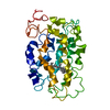
| Structure viewer | Molecule: MolmilJmol/JSmol |
|---|
- Downloads & links
-Download
| PDBx/mmCIF format | 2j4y.cif.gz | 143 KB | Display | PDBx/mmCIF format |
|---|---|---|---|---|
| PDB format | pdb2j4y.ent.gz | 111.8 KB | Display | PDB format |
| PDBx/mmJSON format | 2j4y.json.gz | Tree view | PDBx/mmJSON format | |
| Others |  Other downloads Other downloads |
-Validation report
| Arichive directory | https://data.pdbj.org/pub/pdb/validation_reports/j4/2j4yftp://data.pdbj.org/pub/pdb/validation_reports/j4/2j4y | HTTPS FTP |
|---|
-Related structure data
| Related structure data | 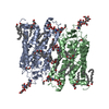 1gzmS S: Starting model for refinement |
|---|---|
| Similar structure data |


-Links
 PDBj
PDBj


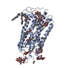
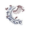
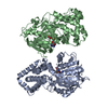
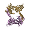
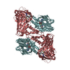
- Assembly
| Deposited unit | 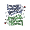
| ||||||||
|---|---|---|---|---|---|---|---|---|---|
| 1 |
| ||||||||
| Unit cell |
| ||||||||
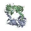
| Noncrystallographic symmetry (NCS) | NCS oper: (Code: given Matrix: (-1, 0.00057, -0.00035), Vector : Details | THE DIMER DESCRIBED HERE IN AN ARTIFACT OF CRYSTALLIZATION AND DOES NOT REPRESENT THE NATURAL OLIGOMERIC STATE OF THE PROTEIN | |
-Components
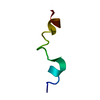
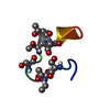
| #1: Protein | Mass: 39034.586 Da / Num. of mol.: 2 / Mutation: YES Source method: isolated from a genetically manipulated source Source: (gene. exp.) BOS TAURUS (cattle) / Tissue: RETINA / Cell: ROD PHOTORECEPTOR / Organ: EYE / Cell line (production host): COS-1 MONKEY KIDNEY CELLS / Production host: CHLOROCEBUS AETHIOPS (grivet) / References: UniProt: P02699#2: Chemical | Retinal  Mass: 284.436 Da / Num. of mol.: 2 / Source method: obtained synthetically / Formula: C20H28O Mass: 284.436 Da / Num. of mol.: 2 / Source method: obtained synthetically / Formula: C20H28O#3: Sugar | N-Acetylglucosamine  Type: D-saccharide, beta linking / Mass: 221.208 Da / Num. of mol.: 2 Type: D-saccharide, beta linking / Mass: 221.208 Da / Num. of mol.: 2Source method: isolated from a genetically manipulated source Formula: C8H15NO6 Compound details | ENGINEERED RESIDUE IN CHAIN A, ASN 2 TO CYS ENGINEERED RESIDUE IN CHAIN A, ASP 282 TO CYS ...ENGINEERED | |
|---|
-Experimental details
-Experiment
| Experiment | Method: X-RAY DIFFRACTION / Number of used crystals: 1 |
|---|
- Sample preparation
| Crystal | Density Matthews: 3.64 Å3/Da / Density % sol: 58 % Description: DATA WAS COLLECTED ON 10 POSITION ALONG A MICROCRYSTALLINE NEEDLE OF 5X5X90 MICRONS |
|---|---|
| Crystal grow | Method: vapor diffusion, sitting drop / pH: 7.5 Details: VAPOUR DIFFUSION IN SITTING DROPS OF 10-15 MG/ML PROTEIN AND 0.2% C8E4,0.05%LDAO AGAINST 1.1-1.7M LITHIUM SULFATE, 0.1M HEPES PH 7.5 |
-Data collection
| Diffraction | Mean temperature: 100 K |
|---|---|
| Diffraction source | Source: SYNCHROTRON / Site: ESRF  / Beamline: ID13 / Wavelength: 0.976 / Beamline: ID13 / Wavelength: 0.976 |
| Detector | Type: MARRESEARCH / Detector: CCD / Date: Jun 18, 2004 / Details: MICROFOCUSING |
| Radiation | Protocol: SINGLE WAVELENGTH / Monochromatic (M) / Laue (L): M / Scattering type: x-ray |
| Radiation wavelength | Wavelength: 0.976 Å / Relative weight: 1 |
| Reflection | Resolution: 3.4→50 Å / Num. obs: 13689 / % possible obs: 95.9 % / Observed criterion σ(I): -3 / Redundancy: 3.03 % / Biso Wilson estimate: 58.2 Å2 / Rmerge(I) obs: 0.24 / Net I/σ(I): 5.42 |
| Reflection shell | Resolution: 3.4→3.5 Å / Redundancy: 2.79 % / Rmerge(I) obs: 0.7 / Mean I/σ(I) obs: 1.79 / % possible all: 84.8 |
- Processing
| Software |
| ||||||||||||||||||||||||||||||||||||||||||||||||||||||||||||||||||||||||||||||||
|---|---|---|---|---|---|---|---|---|---|---|---|---|---|---|---|---|---|---|---|---|---|---|---|---|---|---|---|---|---|---|---|---|---|---|---|---|---|---|---|---|---|---|---|---|---|---|---|---|---|---|---|---|---|---|---|---|---|---|---|---|---|---|---|---|---|---|---|---|---|---|---|---|---|---|---|---|---|---|---|---|---|
| Refinement | Method to determine structure: MOLECULAR REPLACEMENT Starting model: PDB ENTRY 1GZM Resolution: 3.4→50 Å / Rfactor Rfree error: 0.016 / Isotropic thermal model: RESTRAINED / Cross valid method: THROUGHOUT / σ(F): 0 / Stereochemistry target values: MAXIMUM LIKELIHOOD
| ||||||||||||||||||||||||||||||||||||||||||||||||||||||||||||||||||||||||||||||||
| Solvent computation | Bsol: 64.4913 Å2 / ksol: 0.439284 e/Å3 | ||||||||||||||||||||||||||||||||||||||||||||||||||||||||||||||||||||||||||||||||
| Displacement parameters | Biso mean: 55.6579 Å2
| ||||||||||||||||||||||||||||||||||||||||||||||||||||||||||||||||||||||||||||||||
| Refine analyze |
| ||||||||||||||||||||||||||||||||||||||||||||||||||||||||||||||||||||||||||||||||
| Refinement step | Cycle: LAST / Resolution: 3.4→50 Å
| ||||||||||||||||||||||||||||||||||||||||||||||||||||||||||||||||||||||||||||||||
| Refine LS restraints |
| ||||||||||||||||||||||||||||||||||||||||||||||||||||||||||||||||||||||||||||||||
| Refine LS restraints NCS | Rms dev Biso : 1 Å2 / Rms dev position: 0.042 Å / Weight Biso : 10 / Weight position: 100 | ||||||||||||||||||||||||||||||||||||||||||||||||||||||||||||||||||||||||||||||||
| LS refinement shell | Resolution: 3.4→3.55 Å / Total num. of bins used: 8
| ||||||||||||||||||||||||||||||||||||||||||||||||||||||||||||||||||||||||||||||||
| Xplor file |
|
