 Movie
Movie Controller

[English] 日本語
 Yorodumi
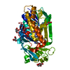
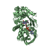
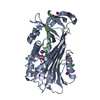
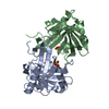
- PDB-7dmb: Crystal structure of trans-methyltransferase CalH complex with SAH -
+ Open data

- Basic information
| Entry | Database: PDB / ID: 7dmb | ||||||
|---|---|---|---|---|---|---|---|
| Title | Crystal structure of trans-methyltransferase CalH complex with SAH | ||||||
 Components Components | Putative methyltransferase | ||||||
 Keywords Keywords | TRANSFERASE / methyltransferase / CalH / Calbistrin A Biosynthesis | ||||||
| Function / homology | : / Methyltransferase type 12 / Methyltransferase domain / methyltransferase activity / methylation / S-adenosyl-L-methionine-dependent methyltransferase superfamily / S-ADENOSYL-L-HOMOCYSTEINE / Putative methyltransferase Function and homology information Function and homology information | ||||||
| Biological species |  | ||||||
| Method |  X-RAY DIFFRACTION / SYNCHROTRON / SAD / Resolution: 2.1 Å X-RAY DIFFRACTION / SYNCHROTRON / SAD / Resolution: 2.1 Å | ||||||
 Authors Authors | Tao, H. / Mori, T. / Abe, I. | ||||||
 Citation Citation | Journal: Angew.Chem.Int.Ed.Engl. / Year: 2021 Title: One Polyketide Synthase, Two Distinct Products: Trans-Acting Enzyme-Controlled Product Divergence in Calbistrin Biosynthesis. Authors: Tao, H. / Mori, T. / Wei, X. / Matsuda, Y. / Abe, I. | ||||||
| History |
|
- Structure visualization
| Structure viewer | Molecule: MolmilJmol/JSmol |
|---|
- Downloads & links
-Download
| PDBx/mmCIF format | 7dmb.cif.gz | 168.8 KB | Display | PDBx/mmCIF format |
|---|---|---|---|---|
| PDB format | pdb7dmb.ent.gz | 128.1 KB | Display | PDB format |
| PDBx/mmJSON format | 7dmb.json.gz | Tree view | PDBx/mmJSON format | |
| Others |  Other downloads Other downloads |
-Validation report
| Arichive directory | https://data.pdbj.org/pub/pdb/validation_reports/dm/7dmbftp://data.pdbj.org/pub/pdb/validation_reports/dm/7dmb | HTTPS FTP |
|---|
-Related structure data
| Similar structure data |
|---|


-Links
 PDBj
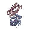
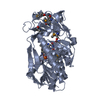
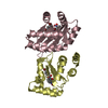
PDBj- Assembly
| Deposited unit | 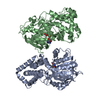
| ||||||||||||
|---|---|---|---|---|---|---|---|---|---|---|---|---|---|
| 1 | 
| ||||||||||||
| 2 | 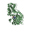
| ||||||||||||
| Unit cell |
|
-Components
| #1: Protein | Mass: 43444.129 Da / Num. of mol.: 2 Source method: isolated from a genetically manipulated source Source: (gene. exp.)  #2: Chemical |   Type: L-peptide linking / Mass: 384.411 Da / Num. of mol.: 2 / Source method: obtained synthetically / Formula: C14H20N6O5S / Feature type: SUBJECT OF INVESTIGATION Type: L-peptide linking / Mass: 384.411 Da / Num. of mol.: 2 / Source method: obtained synthetically / Formula: C14H20N6O5S / Feature type: SUBJECT OF INVESTIGATION#3: Water | ChemComp-HOH / |  Mass: 18.015 Da / Num. of mol.: 501 / Source method: isolated from a natural source / Formula: H2O Mass: 18.015 Da / Num. of mol.: 501 / Source method: isolated from a natural source / Formula: H2OHas ligand of interest | Y | |
|---|
-Experimental details
-Experiment
| Experiment | Method: X-RAY DIFFRACTION / Number of used crystals: 1 |
|---|
- Sample preparation
| Crystal | Density Matthews: 2.07 Å3/Da / Density % sol: 40.64 % |
|---|---|
| Crystal grow | Temperature: 283 K / Method: vapor diffusion, sitting drop / Details: 100 mM HEPES pH 7.0, 20% PEG4000, 0.5 mM SAH |
-Data collection
| Diffraction | Mean temperature: 100 K / Serial crystal experiment: N |
|---|---|
| Diffraction source | Source: SYNCHROTRON / Site: Photon Factory  / Beamline: BL-1A / Wavelength: 1.08 Å / Beamline: BL-1A / Wavelength: 1.08 Å |
| Detector | Type: DECTRIS EIGER X 4M / Detector: PIXEL / Date: Nov 27, 2020 |
| Radiation | Protocol: SINGLE WAVELENGTH / Monochromatic (M) / Laue (L): M / Scattering type: x-ray |
| Radiation wavelength | Wavelength: 1.08 Å / Relative weight: 1 |
| Reflection | Resolution: 2.1→46.99 Å / Num. obs: 39461 / % possible obs: 97 % / Redundancy: 3.5 % / Biso Wilson estimate: 17.22 Å2 / CC1/2: 0.987 / Rmerge(I) obs: 0.131 / Net I/σ(I): 5.3 |
| Reflection shell | Resolution: 2.1→2.16 Å / Redundancy: 3.7 % / Rmerge(I) obs: 0.565 / Mean I/σ(I) obs: 1.6 / Num. unique obs: 3199 / CC1/2: 0.717 |
- Processing
| Software |
| |||||||||||||||||||||||||||||||||||||||||||||||||||||||||||||||||||||||||||||||||||||||||||||||||||||||||
|---|---|---|---|---|---|---|---|---|---|---|---|---|---|---|---|---|---|---|---|---|---|---|---|---|---|---|---|---|---|---|---|---|---|---|---|---|---|---|---|---|---|---|---|---|---|---|---|---|---|---|---|---|---|---|---|---|---|---|---|---|---|---|---|---|---|---|---|---|---|---|---|---|---|---|---|---|---|---|---|---|---|---|---|---|---|---|---|---|---|---|---|---|---|---|---|---|---|---|---|---|---|---|---|---|---|---|
| Refinement | Method to determine structure: SAD / Resolution: 2.1→46.99 Å / SU ML: 0.2505 / Cross valid method: FREE R-VALUE / σ(F): 1.83 / Phase error: 23.3194 Stereochemistry target values: GeoStd + Monomer Library + CDL v1.2
| |||||||||||||||||||||||||||||||||||||||||||||||||||||||||||||||||||||||||||||||||||||||||||||||||||||||||
| Solvent computation | Shrinkage radii: 0.9 Å / VDW probe radii: 1.11 Å / Solvent model: FLAT BULK SOLVENT MODEL | |||||||||||||||||||||||||||||||||||||||||||||||||||||||||||||||||||||||||||||||||||||||||||||||||||||||||
| Displacement parameters | Biso mean: 19.43 Å2 | |||||||||||||||||||||||||||||||||||||||||||||||||||||||||||||||||||||||||||||||||||||||||||||||||||||||||
| Refinement step | Cycle: LAST / Resolution: 2.1→46.99 Å
| |||||||||||||||||||||||||||||||||||||||||||||||||||||||||||||||||||||||||||||||||||||||||||||||||||||||||
| Refine LS restraints |
| |||||||||||||||||||||||||||||||||||||||||||||||||||||||||||||||||||||||||||||||||||||||||||||||||||||||||
| LS refinement shell |
|
