 Movie
Movie Controller


+ Open data

- Basic information
| Entry | Database: PDB / ID: 6a5a | ||||||
|---|---|---|---|---|---|---|---|
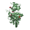
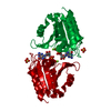
| Title | Crystal structure of plant Receptor-like Kinase ANX1 | ||||||
 Components Components | Receptor-like protein kinase ANXUR1 | ||||||
 Keywords Keywords | TRANSFERASE / a plant receptor | ||||||
| Function / homology |  Function and homology information Function and homology informationpollen tube tip / transmembrane receptor protein tyrosine kinase activity / protein kinase activity / non-specific serine/threonine protein kinase / apical plasma membrane / protein serine kinase activity / protein serine/threonine kinase activity / extracellular region / ATP binding / plasma membrane Similarity search - Function | ||||||
| Biological species |  | ||||||
| Method |  X-RAY DIFFRACTION / SYNCHROTRON / MOLECULAR REPLACEMENT / Resolution: 2.942 Å X-RAY DIFFRACTION / SYNCHROTRON / MOLECULAR REPLACEMENT / Resolution: 2.942 Å | ||||||
 Authors Authors | Xiao, Y. / Chai, J. | ||||||
 Citation Citation | Journal: Nature / Year: 2019 Title: Mechanisms of RALF peptide perception by a heterotypic receptor complex. Authors: Xiao, Y. / Stegmann, M. / Han, Z. / DeFalco, T.A. / Parys, K. / Xu, L. / Belkhadir, Y. / Zipfel, C. / Chai, J. | ||||||
| History |
|
- Structure visualization
| Structure viewer | Molecule: MolmilJmol/JSmol |
|---|
- Downloads & links
-Download
| PDBx/mmCIF format | 6a5a.cif.gz | 156.4 KB | Display | PDBx/mmCIF format |
|---|---|---|---|---|
| PDB format | pdb6a5a.ent.gz | 122.3 KB | Display | PDB format |
| PDBx/mmJSON format | 6a5a.json.gz | Tree view | PDBx/mmJSON format | |
| Others |  Other downloads Other downloads |
-Validation report
| Arichive directory | https://data.pdbj.org/pub/pdb/validation_reports/a5/6a5aftp://data.pdbj.org/pub/pdb/validation_reports/a5/6a5a | HTTPS FTP |
|---|
-Related structure data
| Related structure data |  6a5bC 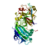 6a5cSC  6a5dC 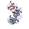 6a5eC S: Starting model for refinement C: citing same article ( |
|---|---|
| Similar structure data |


-Links
 PDBj
PDBj- Assembly
| Deposited unit | 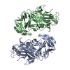
| |||||||||||||||||||||
|---|---|---|---|---|---|---|---|---|---|---|---|---|---|---|---|---|---|---|---|---|---|---|
| 1 | 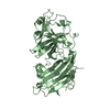
| |||||||||||||||||||||
| 2 | 
| |||||||||||||||||||||
| Unit cell |
| |||||||||||||||||||||
| Noncrystallographic symmetry (NCS) | NCS domain:
NCS domain segments: Component-ID: 1 / Ens-ID: 1 / Beg auth comp-ID: GLY / Beg label comp-ID: GLY / End auth comp-ID: PRO / End label comp-ID: PRO / Auth seq-ID: 26 - 408 / Label seq-ID: 1 - 383
|
-Components
| #1: Protein | Mass: 42893.000 Da / Num. of mol.: 2 Source method: isolated from a genetically manipulated source Source: (gene. exp.) Production host: Insect cell expression vector pTIE1 (others) References: UniProt: Q9SR05, non-specific serine/threonine protein kinase |
|---|
-Experimental details
-Experiment
| Experiment | Method: X-RAY DIFFRACTION / Number of used crystals: 1 |
|---|
- Sample preparation
| Crystal | Density Matthews: 2.71 Å3/Da / Density % sol: 54.55 % |
|---|---|
| Crystal grow | Temperature: 291 K / Method: vapor diffusion, hanging drop Details: 0.2 M ammonium acetate, 0.1 M BIS-Tris pH 6.5, 25%(w/v) Polyethylene glycol 3350 |
-Data collection
| Diffraction | Mean temperature: 100 K |
|---|---|
| Diffraction source | Source: SYNCHROTRON / Site: SSRF  / Beamline: BL17U1 / Wavelength: 1 Å / Beamline: BL17U1 / Wavelength: 1 Å |
| Detector | Type: MAR scanner 345 mm plate / Detector: IMAGE PLATE / Date: Jan 1, 2017 |
| Radiation | Protocol: SINGLE WAVELENGTH / Monochromatic (M) / Laue (L): M / Scattering type: x-ray |
| Radiation wavelength | Wavelength: 1 Å / Relative weight: 1 |
| Reflection | Resolution: 2.94→50 Å / Num. obs: 19284 / % possible obs: 98.3 % / Redundancy: 3.5 % / Rsym value: 0.144 / Net I/σ(I): 17.2 |
| Reflection shell | Resolution: 2.94→3.1 Å / Num. unique obs: 1750 / Rsym value: 0.481 |
- Processing
| Software |
| |||||||||||||||||||||
|---|---|---|---|---|---|---|---|---|---|---|---|---|---|---|---|---|---|---|---|---|---|---|
| Refinement | Method to determine structure: MOLECULAR REPLACEMENT Starting model: 6A5C Resolution: 2.942→41.86 Å / SU ML: 0.36 / Cross valid method: THROUGHOUT / σ(F): 1.34 / Phase error: 27.99 / Stereochemistry target values: ML Details: SF FILE CONTAINS FRIEDEL PAIRS UNDER I/F_MINUS AND I/F_PLUS COLUMNS.
| |||||||||||||||||||||
| Solvent computation | Shrinkage radii: 0.9 Å / VDW probe radii: 1.11 Å / Solvent model: FLAT BULK SOLVENT MODEL | |||||||||||||||||||||
| Displacement parameters | Biso max: 85.39 Å2 / Biso mean: 29.8973 Å2 / Biso min: 8.32 Å2 | |||||||||||||||||||||
| Refinement step | Cycle: final / Resolution: 2.942→41.86 Å
| |||||||||||||||||||||
| Refine LS restraints NCS |
| |||||||||||||||||||||
| LS refinement shell | Resolution: 2.942→3.047 Å
|
