 Movie
Movie Controller

[English] 日本語
 Yorodumi
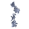
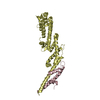
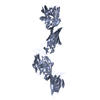
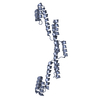
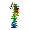
- PDB-1wio: STRUCTURE OF T-CELL SURFACE GLYCOPROTEIN CD4, TETRAGONAL CRYSTAL FORM -
+ Open data

- Basic information
| Entry | Database: PDB / ID: 1wio | ||||||
|---|---|---|---|---|---|---|---|
| Title | STRUCTURE OF T-CELL SURFACE GLYCOPROTEIN CD4, TETRAGONAL CRYSTAL FORM | ||||||
 Components Components | T-CELL SURFACE GLYCOPROTEIN CD4 | ||||||
 Keywords Keywords | GLYCOPROTEIN / IMMUNOGLOBULIN FOLD / TRANSMEMBRANE / T-CELL / MHC LIPOPROTEIN | ||||||
| Function / homology |  Function and homology information Function and homology informationhelper T cell enhancement of adaptive immune response / interleukin-16 binding / interleukin-16 receptor activity / response to methamphetamine hydrochloride / maintenance of protein location in cell / cellular response to ionomycin / T cell selection / MHC class II protein binding / positive regulation of kinase activity / cellular response to granulocyte macrophage colony-stimulating factor stimulus ...helper T cell enhancement of adaptive immune response / interleukin-16 binding / interleukin-16 receptor activity / response to methamphetamine hydrochloride / maintenance of protein location in cell / cellular response to ionomycin / T cell selection / MHC class II protein binding / positive regulation of kinase activity / cellular response to granulocyte macrophage colony-stimulating factor stimulus / interleukin-15-mediated signaling pathway / positive regulation of monocyte differentiation / Nef Mediated CD4 Down-regulation / Alpha-defensins / response to vitamin D / regulation of T cell activation / extracellular matrix structural constituent / Other interleukin signaling / T cell receptor complex / enzyme-linked receptor protein signaling pathway / Translocation of ZAP-70 to Immunological synapse / Phosphorylation of CD3 and TCR zeta chains / positive regulation of protein kinase activity / regulation of calcium ion transport / positive regulation of calcium ion transport into cytosol / macrophage differentiation / Generation of second messenger molecules / immunoglobulin binding / T cell differentiation / Co-inhibition by PD-1 / Binding and entry of HIV virion / coreceptor activity / positive regulation of calcium-mediated signaling / positive regulation of interleukin-2 production / positive regulation of T cell proliferation / cell surface receptor protein tyrosine kinase signaling pathway / protein tyrosine kinase binding / Vpu mediated degradation of CD4 / clathrin-coated endocytic vesicle membrane / calcium-mediated signaling / positive regulation of protein phosphorylation / transmembrane signaling receptor activity / MHC class II protein complex binding / response to estradiol / Downstream TCR signaling / Cargo recognition for clathrin-mediated endocytosis / signaling receptor activity / Clathrin-mediated endocytosis / virus receptor activity / response to ethanol / defense response to Gram-negative bacterium / adaptive immune response / positive regulation of viral entry into host cell / early endosome / cell surface receptor signaling pathway / positive regulation of canonical NF-kappaB signal transduction / positive regulation of ERK1 and ERK2 cascade / cell adhesion / positive regulation of MAPK cascade / immune response / membrane raft / endoplasmic reticulum lumen / external side of plasma membrane / symbiont entry into host cell / lipid binding / protein kinase binding / endoplasmic reticulum membrane / positive regulation of DNA-templated transcription / enzyme binding / signal transduction / protein homodimerization activity / zinc ion binding / identical protein binding / plasma membrane Similarity search - Function | ||||||
| Biological species |  Homo sapiens (human) Homo sapiens (human) | ||||||
| Method |  X-RAY DIFFRACTION / SYNCHROTRON / MOLECULAR REPLACEMENT PLUS ANOMALOUS DIFFRACTION. / Resolution: 3.9 Å X-RAY DIFFRACTION / SYNCHROTRON / MOLECULAR REPLACEMENT PLUS ANOMALOUS DIFFRACTION. / Resolution: 3.9 Å | ||||||
 Authors Authors | Wu, H. / Kwong, P.D. / Hendrickson, W.A. | ||||||
 Citation Citation | Journal: Nature / Year: 1997 Title: Dimeric association and segmental variability in the structure of human CD4. Authors: Wu, H. / Kwong, P.D. / Hendrickson, W.A. | ||||||
| History |
|
- Structure visualization
| Structure viewer | Molecule: MolmilJmol/JSmol |
|---|
- Downloads & links
-Download
| PDBx/mmCIF format | 1wio.cif.gz | 131 KB | Display | PDBx/mmCIF format |
|---|---|---|---|---|
| PDB format | pdb1wio.ent.gz | 100.6 KB | Display | PDB format |
| PDBx/mmJSON format | 1wio.json.gz | Tree view | PDBx/mmJSON format | |
| Others |  Other downloads Other downloads |
-Validation report
| Arichive directory | https://data.pdbj.org/pub/pdb/validation_reports/wi/1wioftp://data.pdbj.org/pub/pdb/validation_reports/wi/1wio | HTTPS FTP |
|---|
-Related structure data


-Links
 PDBj
PDBj


- Assembly
| Deposited unit | 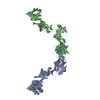
| ||||||||
|---|---|---|---|---|---|---|---|---|---|
| 1 | 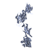
| ||||||||
| 2 | 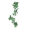
| ||||||||
| Unit cell |
| ||||||||
| Details | THERE ARE TWO MOLECULES PER CRYSTALLOGRAPHIC ASYMMETRIC UNIT. EACH MOLECULE CONTAINS RESIDUES 1 - 363. |
-Components
| #1: Protein | Mass: 40455.477 Da / Num. of mol.: 2 / Fragment: EXTRACELLULAR FRAGMENT / Source method: isolated from a natural source / Details: TETRAGONAL CRYSTAL FORM / Source: (natural) Homo sapiens (human) / References: UniProt: P01730Has protein modification | Y | |
|---|
-Experimental details
-Experiment
| Experiment | Method: X-RAY DIFFRACTION / Number of used crystals: 1 |
|---|
- Sample preparation
| Crystal | Density Matthews: 5.52 Å3/Da / Density % sol: 77.72 % | ||||||||||||||||||||||||||||||||||||||||||||||||||||||||||||
|---|---|---|---|---|---|---|---|---|---|---|---|---|---|---|---|---|---|---|---|---|---|---|---|---|---|---|---|---|---|---|---|---|---|---|---|---|---|---|---|---|---|---|---|---|---|---|---|---|---|---|---|---|---|---|---|---|---|---|---|---|---|
| Crystal grow | *PLUS Temperature: 20 ℃ / pH: 8 / Method: vapor diffusion, hanging dropDetails: Kwong, P.D., (1990) Proc. Natl. Acad. Sci. U.S.A., 87, 6423. | ||||||||||||||||||||||||||||||||||||||||||||||||||||||||||||
| Components of the solutions | *PLUS
|
-Data collection
| Diffraction | Mean temperature: 100 K |
|---|---|
| Diffraction source | Source: SYNCHROTRON / Site: NSLS  / Beamline: X25 / Wavelength: 0.9792 / Beamline: X25 / Wavelength: 0.9792 |
| Detector | Type: FUJI / Detector: IMAGE PLATE |
| Radiation | Monochromatic (M) / Laue (L): M / Scattering type: x-ray |
| Radiation wavelength | Wavelength: 0.9792 Å / Relative weight: 1 |
| Reflection | *PLUS Highest resolution: 3.9 Å / Lowest resolution: 8 Å / Num. obs: 11993 / % possible obs: 80 % / Rmerge(I) obs: 0.112 |
| Reflection shell | *PLUS % possible obs: 53.8 % / Rmerge(I) obs: 0.34 |
- Processing
| Software |
| ||||||||||||||||||||||||||||||||||||||||||||||||||||||||||||
|---|---|---|---|---|---|---|---|---|---|---|---|---|---|---|---|---|---|---|---|---|---|---|---|---|---|---|---|---|---|---|---|---|---|---|---|---|---|---|---|---|---|---|---|---|---|---|---|---|---|---|---|---|---|---|---|---|---|---|---|---|---|
| Refinement | Method to determine structure: MOLECULAR REPLACEMENT PLUS ANOMALOUS DIFFRACTION. Resolution: 3.9→8 Å / σ(F): 2
| ||||||||||||||||||||||||||||||||||||||||||||||||||||||||||||
| Refinement step | Cycle: LAST / Resolution: 3.9→8 Å
| ||||||||||||||||||||||||||||||||||||||||||||||||||||||||||||
| Refine LS restraints |
| ||||||||||||||||||||||||||||||||||||||||||||||||||||||||||||
| Software | *PLUS Name: X-PLOR / Classification: refinement | ||||||||||||||||||||||||||||||||||||||||||||||||||||||||||||
| Refinement | *PLUS Rfactor obs: 0.24 / Rfactor Rwork: 0.24 | ||||||||||||||||||||||||||||||||||||||||||||||||||||||||||||
| Solvent computation | *PLUS | ||||||||||||||||||||||||||||||||||||||||||||||||||||||||||||
| Displacement parameters | *PLUS |
