Resolution: 1.85→1.95 Å / Redundancy: 7.1 % / Rmerge(I) obs: 0.502 / Mean I/σ(I) obs: 3 / % possible all: 96.1
-
Processing
Software
Name
Version
Classification
MxCuBE
datacollection
SHARP
phasing
PHENIX
refine
refinement
MOSFLM
datareduction
SCALA
datascaling
Refinement
Method to determine structure: SAD / Resolution: 1.85→34.63 Å / Cor.coef. Fo:Fc: 0.959 / Cor.coef. Fo:Fc free: 0.936 / SU B: 3.949 / SU ML: 0.117 / Cross valid method: THROUGHOUT / σ(F): 0 / ESU R: 0.163 / ESU R Free: 0.153 / Stereochemistry target values: MAXIMUM LIKELIHOOD / Details: HYDROGENS HAVE BEEN ADDED IN THE RIDING POSITIONS
Rfactor
Num. reflection
% reflection
Selection details
Rfree
0.23692
1328
5 %
RANDOM
Rwork
0.18552
-
-
-
obs
0.1882
25102
96.75 %
-
all
-
26430
-
-
Solvent computation
Ion probe radii: 0.8 Å / Shrinkage radii: 0.8 Å / VDW probe radii: 1.2 Å / Solvent model: MASK
 Movie
Movie Controller


 Open data
 Basic information
 Components
 Keywords
 Function and homology information
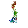
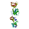
 Streptococcus pneumoniae (bacteria)
 X-RAY DIFFRACTION /
  Authors
 Citation
 Structure visualization
 Downloads & links
 Other downloads


 PDBj
PDBj Assembly
 Mass: 18.015 Da / Num. of mol.: 237 / Source method: isolated from a natural source / Formula: H2O
 Sample preparation
 Processing