- PDB-3h94: Crystal structure of the membrane fusion protein CusB from Escher... -
+
Open data
ID or keywords:
Loading...
-
Basic information
Entry
Database: PDB / ID: 3h94
Title
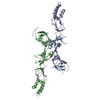
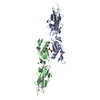
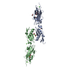
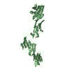
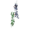
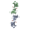
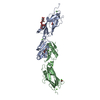
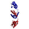
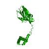
Crystal structure of the membrane fusion protein CusB from Escherichia coli
Components
Cation efflux system protein cusB
Keywords
TRANSPORT PROTEIN / membrane fusion protein / CusCFBA copper/silver efflux system / Copper / Copper transport / Ion transport / Transport
Function / homology
Function and homology information
plasma membrane copper ion transport / copper ion transmembrane transport / response to silver ion / silver ion transmembrane transport / copper ion transmembrane transporter activity / copper ion export / detoxification of copper ion / cell envelope / response to copper ion / transition metal ion binding ...plasma membrane copper ion transport / copper ion transmembrane transport / response to silver ion / silver ion transmembrane transport / copper ion transmembrane transporter activity / copper ion export / detoxification of copper ion / cell envelope / response to copper ion / transition metal ion binding / intracellular copper ion homeostasis / response to toxic substance / outer membrane-bounded periplasmic space / copper ion binding / membrane Similarity search - Function
Helix Hairpins - #730 / : / : / : / CusB, three alpha-helical bundle domain / CusB, barrel-sandwich hybrid domain / : / conserved putative lor/sdh protein from methanococcus maripaludis s2 fold - #20 / : / CusB-like, beta-barrel domain ...Helix Hairpins - #730 / : / : / : / CusB, three alpha-helical bundle domain / CusB, barrel-sandwich hybrid domain / : / conserved putative lor/sdh protein from methanococcus maripaludis s2 fold - #20 / : / CusB-like, beta-barrel domain / conserved putative lor/sdh protein from methanococcus maripaludis s2 fold / Heavy metal binding domain / Heavy metal binding domain / : / Efflux pump adaptor protein, beta barrel domain / RND efflux pump, membrane fusion protein / RNA polymerase II/Efflux pump adaptor protein, barrel-sandwich hybrid domain / Helix Hairpins / Elongation Factor Tu (Ef-tu); domain 3 / Helix non-globular / Special / OB fold (Dihydrolipoamide Acetyltransferase, E2P) / Beta Barrel / Mainly Beta Similarity search - Domain/homology
In the structure databanks used in Yorodumi, some data are registered as the other names, "COVID-19 virus" and "2019-nCoV". Here are the details of the virus and the list of structure data.
Jan 31, 2019. EMDB accession codes are about to change! (news from PDBe EMDB page)
EMDB accession codes are about to change! (news from PDBe EMDB page)
The allocation of 4 digits for EMDB accession codes will soon come to an end. Whilst these codes will remain in use, new EMDB accession codes will include an additional digit and will expand incrementally as the available range of codes is exhausted. The current 4-digit format prefixed with “EMD-” (i.e. EMD-XXXX) will advance to a 5-digit format (i.e. EMD-XXXXX), and so on. It is currently estimated that the 4-digit codes will be depleted around Spring 2019, at which point the 5-digit format will come into force.
The EM Navigator/Yorodumi systems omit the EMD- prefix.
Related info.:Q: What is EMD? / ID/Accession-code notation in Yorodumi/EM Navigator
Yorodumi is a browser for structure data from EMDB, PDB, SASBDB, etc.
This page is also the successor to EM Navigator detail page, and also detail information page/front-end page for Omokage search.
The word "yorodu" (or yorozu) is an old Japanese word meaning "ten thousand". "mi" (miru) is to see.
Related info.:EMDB / PDB / SASBDB / Comparison of 3 databanks / Yorodumi Search / Aug 31, 2016. New EM Navigator & Yorodumi / Yorodumi Papers / Jmol/JSmol / Function and homology information / Changes in new EM Navigator and Yorodumi
 Movie
Movie Controller

 Yorodumi
 Open data
 Basic information
 Components
 Keywords
 Function and homology information
 X-RAY DIFFRACTION /
  Authors
 Citation
 Structure visualization
 Downloads & links
 Other downloads
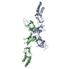

 PDBj
PDBj
 Assembly
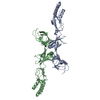
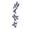

 Mass: 107.868 Da / Num. of mol.: 2 / Source method: obtained synthetically / Formula: Ag
 Sample preparation
 / Beamline: 24-ID-C / Wavelength: 1.3779,0.9792,0.9793,0.9949
 Processing