 Movie
Movie Controller

[English] 日本語
 Yorodumi
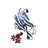
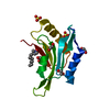
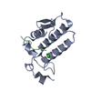
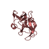
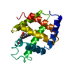
- PDB-1t37: Design of specific inhibitors of phospholipase A2: Crystal struct... -
+ Open data

- Basic information
| Entry | Database: PDB / ID: 1t37 | ||||||
|---|---|---|---|---|---|---|---|
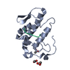
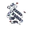
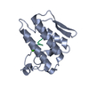
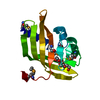
| Title | Design of specific inhibitors of phospholipase A2: Crystal structure of the complex formed between group I phospholipase A2 and a designed pentapeptide Leu-Ala-Ile-Tyr-Ser at 2.6A resolution | ||||||
 Components Components |
| ||||||
 Keywords Keywords |  HYDROLASE / Phospholipase A2 / complex / Inhibition HYDROLASE / Phospholipase A2 / complex / Inhibition | ||||||
| Function / homology |  Function and homology informationphospholipase A2 / phospholipase A2 activity / arachidonic acid secretion / phospholipid metabolic process / lipid catabolic process / calcium ion binding / extracellular region Function and homology informationphospholipase A2 / phospholipase A2 activity / arachidonic acid secretion / phospholipid metabolic process / lipid catabolic process / calcium ion binding / extracellular regionSimilarity search - Function | ||||||
| Biological species |  Naja sagittifera (cobra) Naja sagittifera (cobra) | ||||||
| Method | X-RAY DIFFRACTION / MOLECULAR REPLACEMENT / Resolution: 2.6 Å | ||||||
 Authors Authors | Singh, R.K. / Singh, N. / Jabeen, T. / Makker, J. / Sharma, S. / Dey, S. / Singh, T.P. | ||||||
 Citation Citation | Journal: J.Drug Target. / Year: 2005 Title: Crystal structure of the complex of group I PLA2 with a group II-specific peptide Leu-Ala-Ile-Tyr-Ser (LAIYS) at 2.6 A resolution. Authors: Singh, R.K. / Singh, N. / Jabeen, T. / Sharma, S. / Dey, S. / Singh, T.P. | ||||||
| History |
|
- Structure visualization
| Structure viewer | Molecule: MolmilJmol/JSmol |
|---|
- Downloads & links
-Download
| PDBx/mmCIF format | 1t37.cif.gz | 38.7 KB | Display | PDBx/mmCIF format |
|---|---|---|---|---|
| PDB format | pdb1t37.ent.gz | 26.2 KB | Display | PDB format |
| PDBx/mmJSON format | 1t37.json.gz | Tree view | PDBx/mmJSON format | |
| Others |  Other downloads Other downloads |
-Validation report
| Arichive directory | https://data.pdbj.org/pub/pdb/validation_reports/t3/1t37ftp://data.pdbj.org/pub/pdb/validation_reports/t3/1t37 | HTTPS FTP |
|---|
-Related structure data
| Related structure data |  1zm6C  1lff S: Starting model for refinement C: citing same article ( |
|---|---|
| Similar structure data |


-Links
 PDBj
PDBj

- Assembly
| Deposited unit | 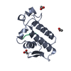
| ||||||||
|---|---|---|---|---|---|---|---|---|---|
| 1 |
| ||||||||
| Unit cell |
|
-Components
| #1: Protein | Mass: 13172.524 Da / Num. of mol.: 1 / Source method: isolated from a natural source / Source: (natural) Naja sagittifera (cobra) / Secretion: Venom / References: UniProt: P60045, phospholipase A2 | ||
|---|---|---|---|
| #2: Protein/peptide | Peptide synthesis Mass: 565.660 Da / Num. of mol.: 1 / Source method: obtained synthetically | ||
| #3: Chemical | Acetate  Mass: 59.044 Da / Num. of mol.: 3 / Source method: obtained synthetically / Formula: C2H3O2 Mass: 59.044 Da / Num. of mol.: 3 / Source method: obtained synthetically / Formula: C2H3O2#4: Water | ChemComp-HOH / | Water Mass: 18.015 Da / Num. of mol.: 55 / Source method: isolated from a natural source / Formula: H2O Mass: 18.015 Da / Num. of mol.: 55 / Source method: isolated from a natural source / Formula: H2O |
-Experimental details
-Experiment
| Experiment | Method: X-RAY DIFFRACTION / Number of used crystals: 1 |
|---|
- Sample preparation
| Crystal | Density Matthews: 2.1 Å3/Da / Density % sol: 43 % |
|---|---|
| Crystal grow | Temperature: 298 K / Method: vapor diffusion, hanging drop / pH: 6 Details: 10mM Sodium phosphate buffer, 35% Ethanol, 2mM Calcium chloride, pH 6.0, VAPOR DIFFUSION, HANGING DROP, temperature 298K |
-Data collection
| Diffraction | Mean temperature: 293 K |
|---|---|
| Diffraction source | Source: ROTATING ANODE / Type: RIGAKU RU300 / Wavelength: 1.5418 Å |
| Detector | Type: MARRESEARCH / Detector: IMAGE PLATE / Date: Jun 4, 2003 / Details: Mirror |
| Radiation | Monochromator: Graphite / Protocol: SINGLE WAVELENGTH / Monochromatic (M) / Laue (L): M / Scattering type: x-ray |
| Radiation wavelength | Wavelength: 1.5418 Å / Relative weight: 1 |
| Reflection | Resolution: 2.6→20 Å / Num. all: 3626 / Num. obs: 3626 / % possible obs: 100 % / Observed criterion σ(F): 0 / Observed criterion σ(I): 0 / Redundancy: 5.6 % / Biso Wilson estimate: 47.7 Å2 / Rsym value: 0.108 / Net I/σ(I): 6.8 |
| Reflection shell | Resolution: 2.6→2.65 Å / Mean I/σ(I) obs: 2 / Rsym value: 0.352 / % possible all: 90.1 |
- Processing
| Software |
| ||||||||||||||||||||||||||||||||||||||||||||||||||||||||||||||||||||||||||||||||
|---|---|---|---|---|---|---|---|---|---|---|---|---|---|---|---|---|---|---|---|---|---|---|---|---|---|---|---|---|---|---|---|---|---|---|---|---|---|---|---|---|---|---|---|---|---|---|---|---|---|---|---|---|---|---|---|---|---|---|---|---|---|---|---|---|---|---|---|---|---|---|---|---|---|---|---|---|---|---|---|---|---|
| Refinement | Method to determine structure: MOLECULAR REPLACEMENT Starting model: 1LFF 1lff Resolution: 2.6→19.36 Å / Rfactor Rfree error: 0.017 / Data cutoff high absF: 721920.77 / Data cutoff low absF: 0 / Isotropic thermal model: RESTRAINED / Cross valid method: THROUGHOUT / σ(F): 0 / Stereochemistry target values: Engh & Huber
| ||||||||||||||||||||||||||||||||||||||||||||||||||||||||||||||||||||||||||||||||
| Solvent computation | Solvent model: FLAT MODEL / Bsol: 90.3398 Å2 / ksol: 0.398971 e/Å3 | ||||||||||||||||||||||||||||||||||||||||||||||||||||||||||||||||||||||||||||||||
| Displacement parameters | Biso mean: 37.3 Å2
| ||||||||||||||||||||||||||||||||||||||||||||||||||||||||||||||||||||||||||||||||
| Refine analyze |
| ||||||||||||||||||||||||||||||||||||||||||||||||||||||||||||||||||||||||||||||||
| Refinement step | Cycle: LAST / Resolution: 2.6→19.36 Å
| ||||||||||||||||||||||||||||||||||||||||||||||||||||||||||||||||||||||||||||||||
| Refine LS restraints |
| ||||||||||||||||||||||||||||||||||||||||||||||||||||||||||||||||||||||||||||||||
| LS refinement shell | Resolution: 2.6→2.76 Å / Rfactor Rfree error: 0.063 / Total num. of bins used: 6
| ||||||||||||||||||||||||||||||||||||||||||||||||||||||||||||||||||||||||||||||||
| Xplor file |
|
