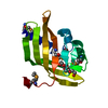
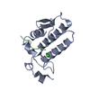
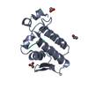
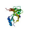
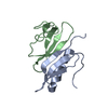
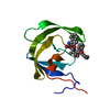
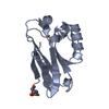
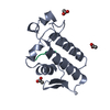
[English] 日本語
Yorodumi
- PDB-3gci: Crystal Structure of the Complex Formed Between a New Isoform of ... -

+
Open data


ID or keywords:

Loading...

-
Basic information

Entry
Database: PDB / ID: 3gci
TitleCrystal Structure of the Complex Formed Between a New Isoform of Phospholipase A2 with C-terminal Amyloid Beta Heptapeptide at 2 A Resolution
Components
  • Heptapeptide from Amyloid beta A4 protein
  • Phospholipase A2 isoform 3
KeywordsHYDROLASE / PHOSPHOLIPASE A2 / HEPTAPEPTIDE / AMYLOID BETA / Lipid degradation / Metal-binding / Secreted
Function / homology
Function and homology information


regulation of epidermal growth factor-activated receptor activity / cytosolic mRNA polyadenylation / collateral sprouting in absence of injury / microglia development / regulation of Wnt signaling pathway / regulation of synapse structure or activity / Formyl peptide receptors bind formyl peptides and many other ligands / axo-dendritic transport / synaptic assembly at neuromuscular junction / signaling receptor activator activity ...regulation of epidermal growth factor-activated receptor activity / cytosolic mRNA polyadenylation / collateral sprouting in absence of injury / microglia development / regulation of Wnt signaling pathway / regulation of synapse structure or activity / Formyl peptide receptors bind formyl peptides and many other ligands / axo-dendritic transport / synaptic assembly at neuromuscular junction / signaling receptor activator activity / smooth endoplasmic reticulum calcium ion homeostasis / calcium-dependent phospholipase A2 activity / axon midline choice point recognition / phospholipase A2 / astrocyte activation involved in immune response / regulation of spontaneous synaptic transmission / mating behavior / NMDA selective glutamate receptor signaling pathway / ciliary rootlet / Lysosome Vesicle Biogenesis / PTB domain binding / Golgi-associated vesicle / arachidonic acid secretion / positive regulation of amyloid fibril formation / neuron remodeling / : / Insertion of tail-anchored proteins into the endoplasmic reticulum membrane / Deregulated CDK5 triggers multiple neurodegenerative pathways in Alzheimer's disease models / suckling behavior / nuclear envelope lumen / dendrite development / COPII-coated ER to Golgi transport vesicle / presynaptic active zone / modulation of excitatory postsynaptic potential / TRAF6 mediated NF-kB activation / Advanced glycosylation endproduct receptor signaling / neuromuscular process controlling balance / The NLRP3 inflammasome / regulation of presynapse assembly / transition metal ion binding / negative regulation of long-term synaptic potentiation / regulation of multicellular organism growth / negative regulation of neuron differentiation / intracellular copper ion homeostasis / ECM proteoglycans / smooth endoplasmic reticulum / positive regulation of T cell migration / spindle midzone / phospholipid metabolic process / Purinergic signaling in leishmaniasis infection / positive regulation of calcium-mediated signaling / lipid catabolic process / protein serine/threonine kinase binding / positive regulation of chemokine production / clathrin-coated pit / regulation of peptidyl-tyrosine phosphorylation / forebrain development / Notch signaling pathway / Mitochondrial protein degradation / neuron projection maintenance / positive regulation of G2/M transition of mitotic cell cycle / positive regulation of protein metabolic process / ionotropic glutamate receptor signaling pathway / positive regulation of glycolytic process / cholesterol metabolic process / response to interleukin-1 / positive regulation of mitotic cell cycle / adult locomotory behavior / axonogenesis / extracellular matrix organization / positive regulation of peptidyl-threonine phosphorylation / platelet alpha granule lumen / trans-Golgi network membrane / dendritic shaft / learning / positive regulation of interleukin-1 beta production / locomotory behavior / positive regulation of long-term synaptic potentiation / central nervous system development / endosome lumen / astrocyte activation / positive regulation of JNK cascade / Post-translational protein phosphorylation / synapse organization / microglial cell activation / regulation of long-term neuronal synaptic plasticity / TAK1-dependent IKK and NF-kappa-B activation / visual learning / serine-type endopeptidase inhibitor activity / neuromuscular junction / recycling endosome / phospholipid binding / cognition / neuron cellular homeostasis / Golgi lumen / positive regulation of inflammatory response / positive regulation of non-canonical NF-kappaB signal transduction / endocytosis / cellular response to amyloid-beta / G2/M transition of mitotic cell cycle
Similarity search - Function
Amyloidogenic glycoprotein, copper-binding / Amyloidogenic glycoprotein, copper-binding domain conserved site / Amyloidogenic glycoprotein, copper-binding domain superfamily / Copper-binding of amyloid precursor, CuBD / Amyloid precursor protein (APP) copper-binding (CuBD) domain signature. / Amyloidogenic glycoprotein, amyloid-beta peptide superfamily / Beta-amyloid peptide (beta-APP) / Amyloidogenic glycoprotein, amyloid-beta peptide / Beta-amyloid precursor protein C-terminal / Amyloidogenic glycoprotein, intracellular domain, conserved site ...Amyloidogenic glycoprotein, copper-binding / Amyloidogenic glycoprotein, copper-binding domain conserved site / Amyloidogenic glycoprotein, copper-binding domain superfamily / Copper-binding of amyloid precursor, CuBD / Amyloid precursor protein (APP) copper-binding (CuBD) domain signature. / Amyloidogenic glycoprotein, amyloid-beta peptide superfamily / Beta-amyloid peptide (beta-APP) / Amyloidogenic glycoprotein, amyloid-beta peptide / Beta-amyloid precursor protein C-terminal / Amyloidogenic glycoprotein, intracellular domain, conserved site / Beta-amyloid precursor protein C-terminus / Amyloid precursor protein (APP) intracellular domain signature. / Amyloid precursor protein (APP) E1 domain profile. / Amyloid precursor protein (APP) E2 domain profile. / Amyloidogenic glycoprotein, extracellular / Amyloidogenic glycoprotein, heparin-binding / Amyloidogenic glycoprotein, E2 domain / E2 domain superfamily / Amyloidogenic glycoprotein, heparin-binding domain superfamily / Amyloid A4 N-terminal heparin-binding / E2 domain of amyloid precursor protein / amyloid A4 / Amyloidogenic glycoprotein / Phospholipase A2, aspartic acid active site / Phospholipase A2 aspartic acid active site. / Phospholipase A2 / Phospholipase A2, histidine active site / Phospholipase A2 histidine active site. / Phospholipase A2 / Phospholipase A2 domain / Phospholipase A2 / Phospholipase A2 / Phospholipase A2 domain / Phospholipase A2 domain superfamily / Proteinase inhibitor I2, Kunitz, conserved site / Pancreatic trypsin inhibitor (Kunitz) family signature. / BPTI/Kunitz family of serine protease inhibitors. / Pancreatic trypsin inhibitor Kunitz domain / Kunitz/Bovine pancreatic trypsin inhibitor domain / Pancreatic trypsin inhibitor (Kunitz) family profile. / Pancreatic trypsin inhibitor Kunitz domain superfamily / PH-like domain superfamily / Up-down Bundle / Mainly Alpha
Similarity search - Domain/homology
Amyloid-beta precursor protein / Acidic phospholipase A2 3
Similarity search - Component
Biological speciesNaja sagittifera (cobra)
HOMO SAPIENS (human)
MethodX-RAY DIFFRACTION / MOLECULAR REPLACEMENT / Resolution: 2.04 Å
AuthorsMirza, Z. / Vikram, G. / Singh, N. / Sinha, M. / Bhushan, A. / Sharma, S. / Srinivasan, A. / Kaur, P. / Singh, T.P.
CitationJournal: To be Published
Title: Crystal Structure of the Complex Formed Between a New Isoform of Phospholipase A2 with C-terminal Amyloid Beta Heptapeptide at 2 A Resolution
Authors: Mirza, Z. / Vikram, G. / Singh, N. / Sinha, M. / Bhushan, A. / Sharma, S. / Srinivasan, A. / Kaur, P. / Singh, T.P.
History
DepositionFeb 22, 2009Deposition site: RCSB / Processing site: PDBJ
Revision 1.0Mar 10, 2009Provider: repository / Type: Initial release
Revision 1.1Jul 13, 2011Group: Version format compliance
Revision 1.2Feb 12, 2014Group: Derived calculations
Revision 1.3Nov 1, 2023Group: Data collection / Database references ...Data collection / Database references / Derived calculations / Refinement description
Category: chem_comp_atom / chem_comp_bond ...chem_comp_atom / chem_comp_bond / database_2 / pdbx_initial_refinement_model / pdbx_struct_conn_angle / struct_conn / struct_site
Item: _database_2.pdbx_DOI / _database_2.pdbx_database_accession ..._database_2.pdbx_DOI / _database_2.pdbx_database_accession / _pdbx_struct_conn_angle.ptnr1_auth_asym_id / _pdbx_struct_conn_angle.ptnr1_auth_comp_id / _pdbx_struct_conn_angle.ptnr1_auth_seq_id / _pdbx_struct_conn_angle.ptnr1_label_asym_id / _pdbx_struct_conn_angle.ptnr1_label_atom_id / _pdbx_struct_conn_angle.ptnr1_label_comp_id / _pdbx_struct_conn_angle.ptnr1_label_seq_id / _pdbx_struct_conn_angle.ptnr3_auth_asym_id / _pdbx_struct_conn_angle.ptnr3_auth_comp_id / _pdbx_struct_conn_angle.ptnr3_auth_seq_id / _pdbx_struct_conn_angle.ptnr3_label_asym_id / _pdbx_struct_conn_angle.ptnr3_label_atom_id / _pdbx_struct_conn_angle.ptnr3_label_comp_id / _pdbx_struct_conn_angle.ptnr3_label_seq_id / _pdbx_struct_conn_angle.value / _struct_conn.pdbx_dist_value / _struct_conn.ptnr1_auth_asym_id / _struct_conn.ptnr1_auth_comp_id / _struct_conn.ptnr1_auth_seq_id / _struct_conn.ptnr1_label_asym_id / _struct_conn.ptnr1_label_atom_id / _struct_conn.ptnr1_label_comp_id / _struct_conn.ptnr1_label_seq_id / _struct_conn.ptnr2_auth_asym_id / _struct_conn.ptnr2_auth_comp_id / _struct_conn.ptnr2_auth_seq_id / _struct_conn.ptnr2_label_asym_id / _struct_conn.ptnr2_label_atom_id / _struct_conn.ptnr2_label_comp_id / _struct_conn.ptnr2_label_seq_id / _struct_site.pdbx_auth_asym_id / _struct_site.pdbx_auth_comp_id / _struct_site.pdbx_auth_seq_id

-
Structure visualization

Structure viewerMolecule:
MolmilJmol/JSmol

Downloads & links

-
Assembly

Deposited unit
A: Phospholipase A2 isoform 3
P: Heptapeptide from Amyloid beta A4 protein
hetero molecules


Theoretical massNumber of molelcules
Total (without water)13,7483
Polymers13,7082
Non-polymers401
Water1,67593
1


  • Idetical with deposited unit
  • defined by author&software
TypeNameSymmetry operationNumber
identity operation1_555x,y,z1
Buried area1030 Å2
ΔGint-20 kcal/mol
Surface area6790 Å2
MethodPISA
Unit cell
Length a, b, c (Å)42.679, 42.679, 65.817
Angle α, β, γ (deg.)90.00, 90.00, 90.00
Int Tables number76
Space group name H-MP41

-
Components

#1: Protein Phospholipase A2 isoform 3 / Phosphatidylcholine 2-acylhydrolase


Mass: 13094.520 Da / Num. of mol.: 1 / Source method: isolated from a natural source / Source: (natural) Naja sagittifera (cobra) / References: UniProt: P60045, phospholipase A2
#2: Protein/peptide Heptapeptide from Amyloid beta A4 protein


Mass: 613.747 Da / Num. of mol.: 1 / Fragment: UNP residues 707-713 / Source method: obtained synthetically
Details: THIS PEPTIDE WAS CHEMICALLY SYNTHESIZED. This sequence occurs naturally in humans.
Source: (synth.) HOMO SAPIENS (human) / References: UniProt: P05067
#3: Chemical ChemComp-CA / CALCIUM ION


Mass: 40.078 Da / Num. of mol.: 1 / Source method: obtained synthetically / Formula: Ca
#4: Water ChemComp-HOH / water


Mass: 18.015 Da / Num. of mol.: 93 / Source method: isolated from a natural source / Formula: H2O
Sequence detailsIN CHAIN A, RESIDUE NUMBER 16 IS SIMPLY SKIPPED. THE AUTHORS BELIEVE THAT THE SEQRES IS CORRECT AND ...IN CHAIN A, RESIDUE NUMBER 16 IS SIMPLY SKIPPED. THE AUTHORS BELIEVE THAT THE SEQRES IS CORRECT AND THESE MUTATIONS ARE PRESENT IN THE DEPOSITED SEQUENCE.

-
Experimental details

-
Experiment

ExperimentMethod: X-RAY DIFFRACTION / Number of used crystals: 1

-
Sample preparation

CrystalDensity Matthews: 2.19 Å3/Da / Density % sol: 43.74 %
Crystal growTemperature: 290 K / Method: vapor diffusion, hanging drop / pH: 6
Details: 10mM Sodium phosphate, pH 6.0, 1mM CaCl2, VAPOR DIFFUSION, HANGING DROP, temperature 290K

-
Data collection

DiffractionMean temperature: 300 K
Diffraction sourceSource: ROTATING ANODE / Type: RIGAKU RU300 / Wavelength: 1.5418 Å
DetectorType: MARRESEARCH / Detector: IMAGE PLATE / Date: Sep 23, 2008 / Details: MIRROR
RadiationMonochromator: GRAPHITE / Protocol: SINGLE WAVELENGTH / Monochromatic (M) / Laue (L): M / Scattering type: x-ray
Radiation wavelengthWavelength: 1.5418 Å / Relative weight: 1
ReflectionResolution: 2.04→20 Å / Num. all: 7596 / Num. obs: 7596 / % possible obs: 97.6 % / Observed criterion σ(F): 0 / Observed criterion σ(I): 0 / Rsym value: 0.115 / Net I/σ(I): 7.8
Reflection shellResolution: 2.04→2.09 Å / Mean I/σ(I) obs: 2.1 / Rsym value: 0.305 / % possible all: 96.7

-
Processing

Software
NameVersionClassification
DENZOdata reduction
MOLREPphasing
REFMAC5.2.0019refinement
AUTOMARdata reduction
SCALEPACKdata scaling
RefinementMethod to determine structure: MOLECULAR REPLACEMENT
Starting model: PDB ENTRY 1MF4
Resolution: 2.04→19.51 Å / Cor.coef. Fo:Fc: 0.949 / Cor.coef. Fo:Fc free: 0.926 / SU B: 4.618 / SU ML: 0.128 / Cross valid method: THROUGHOUT / σ(F): 0 / σ(I): 0 / ESU R: 0.232 / ESU R Free: 0.178 / Stereochemistry target values: MAXIMUM LIKELIHOOD / Details: HYDROGENS HAVE BEEN ADDED IN THE RIDING POSITIONS
RfactorNum. reflection% reflectionSelection details
Rfree0.22137 340 4.6 %RANDOM
Rwork0.18436 ---
all0.19 7596 --
obs0.18606 7065 97.91 %-
Solvent computationIon probe radii: 0.8 Å / Shrinkage radii: 0.8 Å / VDW probe radii: 1.2 Å / Solvent model: MASK
Displacement parametersBiso mean: 28.418 Å2
Baniso -1Baniso -2Baniso -3
1--0.43 Å20 Å20 Å2
2---0.43 Å20 Å2
3---0.87 Å2
Refinement stepCycle: LAST / Resolution: 2.04→19.51 Å
ProteinNucleic acidLigandSolventTotal
Num. atoms949 0 1 93 1043
Refine LS restraints
Refine-IDTypeDev idealDev ideal targetNumber
X-RAY DIFFRACTIONr_bond_refined_d0.0120.022972
X-RAY DIFFRACTIONr_angle_refined_deg1.1721.9351319
X-RAY DIFFRACTIONr_dihedral_angle_1_deg4.8845123
X-RAY DIFFRACTIONr_dihedral_angle_2_deg33.772548
X-RAY DIFFRACTIONr_dihedral_angle_3_deg15.61115145
X-RAY DIFFRACTIONr_dihedral_angle_4_deg13.796155
X-RAY DIFFRACTIONr_chiral_restr0.1030.2138
X-RAY DIFFRACTIONr_gen_planes_refined0.0040.02761
X-RAY DIFFRACTIONr_nbd_refined0.2030.2429
X-RAY DIFFRACTIONr_nbtor_refined0.2990.2649
X-RAY DIFFRACTIONr_xyhbond_nbd_refined0.1750.265
X-RAY DIFFRACTIONr_metal_ion_refined0.0130.21
X-RAY DIFFRACTIONr_symmetry_vdw_refined0.2740.228
X-RAY DIFFRACTIONr_symmetry_hbond_refined0.30.217
X-RAY DIFFRACTIONr_mcbond_it0.7321.5636
X-RAY DIFFRACTIONr_mcangle_it1.4082985
X-RAY DIFFRACTIONr_scbond_it1.8783387
X-RAY DIFFRACTIONr_scangle_it2.9444.5334
LS refinement shellResolution: 2.04→2.092 Å / Total num. of bins used: 20
RfactorNum. reflection% reflection
Rfree0.242 28 -
Rwork0.257 493 -
obs--96.66 %

+
About Yorodumi

-
News

-
Feb 9, 2022. New format data for meta-information of EMDB entries

New format data for meta-information of EMDB entries

  • Version 3 of the EMDB header file is now the official format.
  • The previous official version 1.9 will be removed from the archive.

Related info.:EMDB header

External links:wwPDB to switch to version 3 of the EMDB data model

-
Aug 12, 2020. Covid-19 info

Covid-19 info

URL: https://pdbjlvh1.pdbj.org/emnavi/covid19.php

New page: Covid-19 featured information page in EM Navigator.

Related info.:Covid-19 info / Mar 5, 2020. Novel coronavirus structure data

+
Mar 5, 2020. Novel coronavirus structure data

Novel coronavirus structure data

Related info.:Yorodumi Speices / Aug 12, 2020. Covid-19 info

External links:COVID-19 featured content - PDBj / Molecule of the Month (242):Coronavirus Proteases

+
Jan 31, 2019. EMDB accession codes are about to change! (news from PDBe EMDB page)

EMDB accession codes are about to change! (news from PDBe EMDB page)

  • The allocation of 4 digits for EMDB accession codes will soon come to an end. Whilst these codes will remain in use, new EMDB accession codes will include an additional digit and will expand incrementally as the available range of codes is exhausted. The current 4-digit format prefixed with “EMD-” (i.e. EMD-XXXX) will advance to a 5-digit format (i.e. EMD-XXXXX), and so on. It is currently estimated that the 4-digit codes will be depleted around Spring 2019, at which point the 5-digit format will come into force.
  • The EM Navigator/Yorodumi systems omit the EMD- prefix.

Related info.:Q: What is EMD? / ID/Accession-code notation in Yorodumi/EM Navigator

External links:EMDB Accession Codes are Changing Soon! / Contact to PDBj

+
Jul 12, 2017. Major update of PDB

Major update of PDB

  • wwPDB released updated PDB data conforming to the new PDBx/mmCIF dictionary.
  • This is a major update changing the version number from 4 to 5, and with Remediation, in which all the entries are updated.
  • In this update, many items about electron microscopy experimental information are reorganized (e.g. em_software).
  • Now, EM Navigator and Yorodumi are based on the updated data.

External links:wwPDB Remediation / Enriched Model Files Conforming to OneDep Data Standards Now Available in the PDB FTP Archive

-
Yorodumi

Thousand views of thousand structures

  • Yorodumi is a browser for structure data from EMDB, PDB, SASBDB, etc.
  • This page is also the successor to EM Navigator detail page, and also detail information page/front-end page for Omokage search.
  • The word "yorodu" (or yorozu) is an old Japanese word meaning "ten thousand". "mi" (miru) is to see.

Related info.:EMDB / PDB / SASBDB / Comparison of 3 databanks / Yorodumi Search / Aug 31, 2016. New EM Navigator & Yorodumi / Yorodumi Papers / Jmol/JSmol / Function and homology information / Changes in new EM Navigator and Yorodumi

Read more