 Movie
Movie Controller


+ Open data

- Basic information
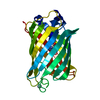
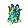
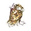
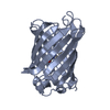
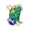
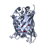
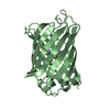
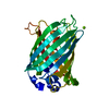
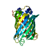
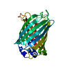
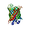
| Entry | Database: PDB / ID: 1q73 | ||||||
|---|---|---|---|---|---|---|---|
| Title | S65T Q80R Y145C T203C Green Fluorescent Protein (GFP) pH 8.5 | ||||||
 Components Components | Green Fluorescent Protein | ||||||
 Keywords Keywords | LUMINESCENT PROTEIN / green fluorescent protein / GFP / fluorophore / chromophore | ||||||
| Function / homology |  Function and homology information Function and homology informationserine-type endopeptidase inhibitor activity / extracellular space / metal ion binding Similarity search - Function | ||||||
| Biological species |   Aequorea victoria (jellyfish) Aequorea victoria (jellyfish) | ||||||
| Method |  X-RAY DIFFRACTION / SYNCHROTRON / MOLECULAR REPLACEMENT / Resolution: 1.6 Å X-RAY DIFFRACTION / SYNCHROTRON / MOLECULAR REPLACEMENT / Resolution: 1.6 Å | ||||||
 Authors Authors | Jain, R.K. / Ranganathan, R. | ||||||
 Citation Citation | Journal: Proc.Natl.Acad.Sci.USA / Year: 2004 Title: Local complexity of amino acid interactions in a protein core. Authors: Jain, R.K. / Ranganathan, R. | ||||||
| History |
|
- Structure visualization
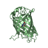
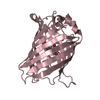
| Structure viewer | Molecule: MolmilJmol/JSmol |
|---|
- Downloads & links
-Download
| PDBx/mmCIF format | 1q73.cif.gz | 63.8 KB | Display | PDBx/mmCIF format |
|---|---|---|---|---|
| PDB format | pdb1q73.ent.gz | 46 KB | Display | PDB format |
| PDBx/mmJSON format | 1q73.json.gz | Tree view | PDBx/mmJSON format | |
| Others |  Other downloads Other downloads |
-Validation report
| Summary document | 1q73_validation.pdf.gz | 429.2 KB | Display | wwPDB validaton report |
|---|---|---|---|---|
| Full document | 1q73_full_validation.pdf.gz | 431.3 KB | Display | |
| Data in XML | 1q73_validation.xml.gz | 13.6 KB | Display | |
| Data in CIF | 1q73_validation.cif.gz | 20 KB | Display | |
| Arichive directory | https://data.pdbj.org/pub/pdb/validation_reports/q7/1q73ftp://data.pdbj.org/pub/pdb/validation_reports/q7/1q73 | HTTPS FTP |
-Related structure data
| Related structure data | 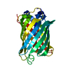 1q4aC 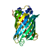 1q4bC 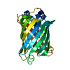 1q4cC  1q4dC  1q4eC  1emaS C: citing same article ( S: Starting model for refinement |
|---|---|
| Similar structure data |


-Links
 PDBj
PDBj


- Assembly
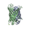
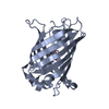
| Deposited unit | 
| ||||||||
|---|---|---|---|---|---|---|---|---|---|
| 1 |
| ||||||||
| Unit cell |
|
-Components
| #1: Protein | Mass: 26887.391 Da / Num. of mol.: 1 / Mutation: Q80R, S65T, Y145C, T203C Source method: isolated from a genetically manipulated source Source: (gene. exp.) Aequorea victoria (jellyfish) / Plasmid: pRSET-B(Invitrogen) / Species (production host): Escherichia coli / Production host:  |
|---|---|
| #2: Water | ChemComp-HOH /  Mass: 18.015 Da / Num. of mol.: 255 / Source method: isolated from a natural source / Formula: H2O Mass: 18.015 Da / Num. of mol.: 255 / Source method: isolated from a natural source / Formula: H2O |
| Sequence details | RESIDUE SER A 65 IS MUTATED TO THR A 65. THR A 65, TYR A 66 AND GLY A 67 ARE MODIFIED TO MAKE ...RESIDUE SER A 65 IS MUTATED TO THR A 65. THR A 65, TYR A 66 AND GLY A 67 ARE MODIFIED TO MAKE CHROMOPHOR |
-Experimental details
-Experiment
| Experiment | Method: X-RAY DIFFRACTION / Number of used crystals: 1 |
|---|
- Sample preparation
| Crystal | Density Matthews: 1.88 Å3/Da / Density % sol: 34.12 % | |||||||||||||||||||||||||||||||||||||||||||||||||
|---|---|---|---|---|---|---|---|---|---|---|---|---|---|---|---|---|---|---|---|---|---|---|---|---|---|---|---|---|---|---|---|---|---|---|---|---|---|---|---|---|---|---|---|---|---|---|---|---|---|---|
| Crystal grow | Temperature: 298 K / Method: vapor diffusion, hanging drop / pH: 8.5 Details: PEG 4000, MgCl2, BME, pH 8.5, VAPOR DIFFUSION, HANGING DROP, temperature 298K | |||||||||||||||||||||||||||||||||||||||||||||||||
| Crystal grow | *PLUS Method: vapor diffusion, hanging drop / PH range low: 8.5 / PH range high: 8.1 | |||||||||||||||||||||||||||||||||||||||||||||||||
| Components of the solutions | *PLUS
|
-Data collection
| Diffraction source | Source: SYNCHROTRON / Site: SSRL  / Beamline: BL7-1 / Beamline: BL7-1 |
|---|---|
| Radiation | Protocol: SINGLE WAVELENGTH / Monochromatic (M) / Laue (L): M / Scattering type: x-ray |
| Radiation wavelength | Relative weight: 1 |
| Reflection | Resolution: 1.6→25 Å / Num. all: 28518 / Num. obs: 28518 / % possible obs: 95.6 % |
| Reflection | *PLUS Redundancy: 3 % / Rmerge(I) obs: 0.078 |
| Reflection shell | *PLUS % possible obs: 90.2 % / Rmerge(I) obs: 0.456 / Mean I/σ(I) obs: 2 |
- Processing
| Software |
| |||||||||||||||||||||||||
|---|---|---|---|---|---|---|---|---|---|---|---|---|---|---|---|---|---|---|---|---|---|---|---|---|---|---|
| Refinement | Method to determine structure: MOLECULAR REPLACEMENT Starting model: PDB ENTRY 1EMA Resolution: 1.6→25 Å / σ(F): 0
| |||||||||||||||||||||||||
| Refinement step | Cycle: LAST / Resolution: 1.6→25 Å
| |||||||||||||||||||||||||
| Refinement | *PLUS Rfactor Rfree: 0.21 | |||||||||||||||||||||||||
| Solvent computation | *PLUS | |||||||||||||||||||||||||
| Displacement parameters | *PLUS | |||||||||||||||||||||||||
| Refine LS restraints | *PLUS
|
