 Movie
Movie Controller


+ Open data

- Basic information
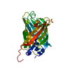
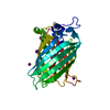
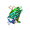
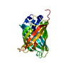
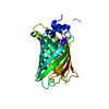
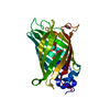
| Entry | Database: PDB / ID: 3ako | |||||||||
|---|---|---|---|---|---|---|---|---|---|---|
| Title | Crystal Structure of the Reassembled Venus | |||||||||
 Components Components | (Venus) x 2 | |||||||||
 Keywords Keywords | FLUORESCENT PROTEIN / Venus / GFP | |||||||||
| Function / homology | HSP40/DNAj peptide-binding domain - #10 / HSP40/DNAj peptide-binding domain / Other non-globular / Green Fluorescent Protein / Green fluorescent protein / Special / Beta Barrel / Mainly Beta / 3,6,9,12,15,18,21-HEPTAOXATRICOSANE-1,23-DIOL Function and homology information Function and homology information | |||||||||
| Biological species | Plant transformation vector pSITEII-4C1 (others) | |||||||||
| Method |  X-RAY DIFFRACTION / SYNCHROTRON / MOLECULAR REPLACEMENT / Resolution: 2.1 Å X-RAY DIFFRACTION / SYNCHROTRON / MOLECULAR REPLACEMENT / Resolution: 2.1 Å | |||||||||
 Authors Authors | Isogai, M. / Tada, T. | |||||||||
 Citation Citation | Journal: Bioorg.Med.Chem.Lett. / Year: 2011 Title: Structure and characteristics of reassembled fluorescent protein, a new insight into the reassembly mechanisms Authors: Isogai, M. / Kawamoto, Y. / Inahata, K. / Fukada, H. / Sugimoto, K. / Tada, T. | |||||||||
| History |
|
- Structure visualization
| Structure viewer | Molecule: MolmilJmol/JSmol |
|---|
- Downloads & links
-Download
| PDBx/mmCIF format | 3ako.cif.gz | 207.6 KB | Display | PDBx/mmCIF format |
|---|---|---|---|---|
| PDB format | pdb3ako.ent.gz | 165.1 KB | Display | PDB format |
| PDBx/mmJSON format | 3ako.json.gz | Tree view | PDBx/mmJSON format | |
| Others |  Other downloads Other downloads |
-Validation report
| Arichive directory | https://data.pdbj.org/pub/pdb/validation_reports/ak/3akoftp://data.pdbj.org/pub/pdb/validation_reports/ak/3ako | HTTPS FTP |
|---|
-Related structure data
| Related structure data | 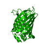 1mywS S: Starting model for refinement |
|---|---|
| Similar structure data |


-Links
 PDBj
PDBj- Assembly
| Deposited unit | 
| ||||||||
|---|---|---|---|---|---|---|---|---|---|
| 1 | 
| ||||||||
| 2 | 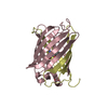
| ||||||||
| 3 | 
| ||||||||
| 4 | 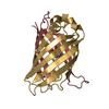
| ||||||||
| Unit cell |
|
-Components
| #1: Protein | Mass: 19895.383 Da / Num. of mol.: 4 / Fragment: N-terminal fragment Source method: isolated from a genetically manipulated source Source: (gene. exp.) Plant transformation vector pSITEII-4C1 (others) Production host:  #2: Protein | Mass: 10651.938 Da / Num. of mol.: 4 / Fragment: C-terminal fragment Source method: isolated from a genetically manipulated source Source: (gene. exp.) Plant transformation vector pSITEII-4C1 (others) Production host: #3: Chemical | ChemComp-SO4 / |   Mass: 96.063 Da / Num. of mol.: 1 / Source method: obtained synthetically / Formula: SO4 Mass: 96.063 Da / Num. of mol.: 1 / Source method: obtained synthetically / Formula: SO4#4: Chemical | ChemComp-PE8 / |   Mass: 370.436 Da / Num. of mol.: 1 / Source method: obtained synthetically / Formula: C16H34O9 Mass: 370.436 Da / Num. of mol.: 1 / Source method: obtained synthetically / Formula: C16H34O9#5: Water | ChemComp-HOH / |  Mass: 18.015 Da / Num. of mol.: 651 / Source method: isolated from a natural source / Formula: H2O Mass: 18.015 Da / Num. of mol.: 651 / Source method: isolated from a natural source / Formula: H2OHas protein modification | Y | Sequence details | SEQUENCE DATABASES OF THE ENTITIES ARE NOT AVAILABLE IN UNIPROT AT PRESENT. SEQUENCE OF ENTITY 1 IS ...SEQUENCE DATABASES OF THE ENTITIES ARE NOT AVAILABLE IN UNIPROT AT PRESENT. SEQUENCE OF ENTITY 1 IS THE SAME AS RESIDUES 1-155 OF GB ADE48838, WITH N-TERMINAL EXPRESSION | |
|---|
-Experimental details
-Experiment
| Experiment | Method: X-RAY DIFFRACTION / Number of used crystals: 1 |
|---|
- Sample preparation
| Crystal | Density Matthews: 2.2 Å3/Da / Density % sol: 44.07 % |
|---|---|
| Crystal grow | Temperature: 293 K / Method: vapor diffusion, sitting drop / pH: 7.5 Details: 25% PEG 3350, 0.1M HEPES, 0.2M lithium sulfate , pH 7.5, VAPOR DIFFUSION, SITTING DROP, temperature 293K |
-Data collection
| Diffraction | Mean temperature: 100 K |
|---|---|
| Diffraction source | Source: SYNCHROTRON / Site: Photon Factory  / Beamline: BL-5A / Wavelength: 1 Å / Beamline: BL-5A / Wavelength: 1 Å |
| Detector | Type: ADSC QUANTUM 315r / Detector: CCD / Date: Apr 28, 2010 |
| Radiation | Monochromator: Numerical link type Si(111) / Protocol: SINGLE WAVELENGTH / Monochromatic (M) / Laue (L): M / Scattering type: x-ray |
| Radiation wavelength | Wavelength: 1 Å / Relative weight: 1 |
| Reflection | Resolution: 2.1→50 Å / Num. all: 63893 / Num. obs: 60532 / % possible obs: 94.7 % / Redundancy: 11 % / Rmerge(I) obs: 0.102 / Net I/σ(I): 23.9 |
| Reflection shell | Resolution: 2.1→2.18 Å / Redundancy: 9.3 % / Rmerge(I) obs: 0.329 / Mean I/σ(I) obs: 5.8 / % possible all: 99.9 |
- Processing
| Software |
| ||||||||||||||||||||||||||||||||||||||||||||||||||||||||||||||||||||||||||||||||||||||||||||||||||||||||||||||||||||||||||||||||||||||||||||||||||||||||||||||||||||||||||
|---|---|---|---|---|---|---|---|---|---|---|---|---|---|---|---|---|---|---|---|---|---|---|---|---|---|---|---|---|---|---|---|---|---|---|---|---|---|---|---|---|---|---|---|---|---|---|---|---|---|---|---|---|---|---|---|---|---|---|---|---|---|---|---|---|---|---|---|---|---|---|---|---|---|---|---|---|---|---|---|---|---|---|---|---|---|---|---|---|---|---|---|---|---|---|---|---|---|---|---|---|---|---|---|---|---|---|---|---|---|---|---|---|---|---|---|---|---|---|---|---|---|---|---|---|---|---|---|---|---|---|---|---|---|---|---|---|---|---|---|---|---|---|---|---|---|---|---|---|---|---|---|---|---|---|---|---|---|---|---|---|---|---|---|---|---|---|---|---|---|---|---|
| Refinement | Method to determine structure: MOLECULAR REPLACEMENT Starting model: PDB ENTRY 1MYW Resolution: 2.1→49.9 Å / Cor.coef. Fo:Fc: 0.948 / Cor.coef. Fo:Fc free: 0.917 / SU B: 4.276 / SU ML: 0.115 / Isotropic thermal model: Isotropic / Cross valid method: THROUGHOUT / ESU R: 0.189 / ESU R Free: 0.178 / Stereochemistry target values: MAXIMUM LIKELIHOOD / Details: HYDROGENS HAVE BEEN ADDED IN THE RIDING POSITIONS
| ||||||||||||||||||||||||||||||||||||||||||||||||||||||||||||||||||||||||||||||||||||||||||||||||||||||||||||||||||||||||||||||||||||||||||||||||||||||||||||||||||||||||||
| Solvent computation | Ion probe radii: 0.8 Å / Shrinkage radii: 0.8 Å / VDW probe radii: 1.4 Å / Solvent model: MASK | ||||||||||||||||||||||||||||||||||||||||||||||||||||||||||||||||||||||||||||||||||||||||||||||||||||||||||||||||||||||||||||||||||||||||||||||||||||||||||||||||||||||||||
| Displacement parameters | Biso mean: 17.411 Å2
| ||||||||||||||||||||||||||||||||||||||||||||||||||||||||||||||||||||||||||||||||||||||||||||||||||||||||||||||||||||||||||||||||||||||||||||||||||||||||||||||||||||||||||
| Refinement step | Cycle: LAST / Resolution: 2.1→49.9 Å
| ||||||||||||||||||||||||||||||||||||||||||||||||||||||||||||||||||||||||||||||||||||||||||||||||||||||||||||||||||||||||||||||||||||||||||||||||||||||||||||||||||||||||||
| Refine LS restraints |
| ||||||||||||||||||||||||||||||||||||||||||||||||||||||||||||||||||||||||||||||||||||||||||||||||||||||||||||||||||||||||||||||||||||||||||||||||||||||||||||||||||||||||||
| LS refinement shell | Resolution: 2.1→2.154 Å / Total num. of bins used: 20
|
