 ムービー
ムービー コントローラー


+ データを開く

- 基本情報
基本情報
| 登録情報 | データベース: PDB / ID: 1n3i | |||||||||
|---|---|---|---|---|---|---|---|---|---|---|
| タイトル | Crystal Structure of Mycobacterium tuberculosis PNP with transition state analog DADMe-ImmH | |||||||||
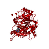
 要素 要素 | Purine Nucleoside Phosphorylase | |||||||||
 キーワード キーワード | TRANSFERASE / transition state complex / trimer / PNP | |||||||||
| 機能・相同性 |  機能・相同性情報 機能・相同性情報deoxyguanosine catabolic process / beta-alanine metabolic process / pantoate-beta-alanine ligase (AMP-forming) / pantoate-beta-alanine ligase activity / pantothenate biosynthetic process / purine-nucleoside phosphorylase / purine-nucleoside phosphorylase activity / manganese ion binding / magnesium ion binding / ATP binding ...deoxyguanosine catabolic process / beta-alanine metabolic process / pantoate-beta-alanine ligase (AMP-forming) / pantoate-beta-alanine ligase activity / pantothenate biosynthetic process / purine-nucleoside phosphorylase / purine-nucleoside phosphorylase activity / manganese ion binding / magnesium ion binding / ATP binding / cytoplasm / cytosol 類似検索 - 分子機能 | |||||||||
| 生物種 |   Mycobacterium tuberculosis (結核菌) Mycobacterium tuberculosis (結核菌) | |||||||||
| 手法 |  X線回折 / フーリエ合成 / 解像度: 1.9 Å X線回折 / フーリエ合成 / 解像度: 1.9 Å | |||||||||
 データ登録者 データ登録者 | Lewandowicz, A. / Shi, W. / Evans, G.B. / Tyler, P.C. / Furneaux, R.H. / Basso, L.A. / Santos, D.S. / Almo, S.C. / Schramm, V.L. | |||||||||
 引用 引用 | ジャーナル: BIOCHEMISTRY / 年: 2003 タイトル: Over-The-Barrier Transition State Analogues Provide New Chemistries for Inhibitor Design: The Case of Purine Nucleoside Phosphorylase 著者: Lewandowicz, A. / Shi, W. / Evans, G.B. / Tyler, P.C. / Furneaux, R.H. / Basso, L.A. / Santos, D.S. / Almo, S.C. / Schramm, V.L. | |||||||||
| 履歴 |
|
- 構造の表示
構造の表示
| 構造ビューア | 分子: MolmilJmol/JSmol |
|---|
- ダウンロードとリンク
-ダウンロード
| PDBx/mmCIF形式 | 1n3i.cif.gz | 158.6 KB | 表示 | PDBx/mmCIF形式 |
|---|---|---|---|---|
| PDB形式 | pdb1n3i.ent.gz | 125.7 KB | 表示 | PDB形式 |
| PDBx/mmJSON形式 | 1n3i.json.gz | ツリー表示 | PDBx/mmJSON形式 | |
| その他 |  その他のダウンロード その他のダウンロード |
-検証レポート
| アーカイブディレクトリ | https://data.pdbj.org/pub/pdb/validation_reports/n3/1n3iftp://data.pdbj.org/pub/pdb/validation_reports/n3/1n3i | HTTPS FTP |
|---|
-関連構造データ
| 関連構造データ | 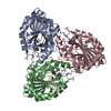 1g2oS S: 精密化の開始モデル |
|---|---|
| 類似構造データ |


-リンク
 PDBj
PDBj- 集合体
集合体
| 登録構造単位 | 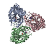
| ||||||||
|---|---|---|---|---|---|---|---|---|---|
| 1 |
| ||||||||
| 単位格子 |
| ||||||||
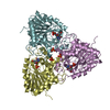
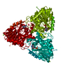
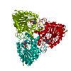
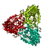
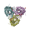
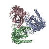
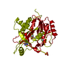
| 詳細 | The biological assembly is the trimer in the asymmetric unit. |
-要素
| #1: タンパク質 | 分子量: 27599.457 Da / 分子数: 3 / 由来タイプ: 組換発現 由来: (組換発現) Mycobacterium tuberculosis (結核菌)プラスミド: PET-23A(+) / 生物種 (発現宿主): Escherichia coli / 発現宿主: 参照: UniProt: P0A538, UniProt: P9WIL5*PLUS, purine-nucleoside phosphorylase #2: 化合物 |   分子量: 94.971 Da / 分子数: 3 / 由来タイプ: 合成 / 式: PO4 分子量: 94.971 Da / 分子数: 3 / 由来タイプ: 合成 / 式: PO4#3: 化合物 | 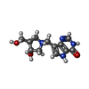  分子量: 265.288 Da / 分子数: 3 / 由来タイプ: 合成 / 式: C12H17N4O3 分子量: 265.288 Da / 分子数: 3 / 由来タイプ: 合成 / 式: C12H17N4O3#4: 水 | ChemComp-HOH / |  分子量: 18.015 Da / 分子数: 432 / 由来タイプ: 天然 / 式: H2O 分子量: 18.015 Da / 分子数: 432 / 由来タイプ: 天然 / 式: H2O |
|---|
-実験情報
-実験
| 実験 | 手法: X線回折 / 使用した結晶の数: 1 |
|---|
- 試料調製
試料調製
| 結晶 | マシュー密度: 2.37 Å3/Da / 溶媒含有率: 48.15 % | |||||||||||||||||||||||||||||||||||||||||||||||||
|---|---|---|---|---|---|---|---|---|---|---|---|---|---|---|---|---|---|---|---|---|---|---|---|---|---|---|---|---|---|---|---|---|---|---|---|---|---|---|---|---|---|---|---|---|---|---|---|---|---|---|
| 結晶化 | 温度: 291 K / 手法: 蒸気拡散法, ハンギングドロップ法 / pH: 8 詳細: PEG4000, Magnesium Chloride, Tris, pH 8.0, VAPOR DIFFUSION, HANGING DROP, temperature 291K | |||||||||||||||||||||||||||||||||||||||||||||||||
| 結晶化 | *PLUS 手法: 蒸気拡散法 | |||||||||||||||||||||||||||||||||||||||||||||||||
| 溶液の組成 | *PLUS
|
-データ収集
| 回折 | 平均測定温度: 100 K |
|---|---|
| 放射光源 | 由来: 回転陽極 / タイプ: RIGAKU / 波長: 1.5418 Å |
| 検出器 | タイプ: RIGAKU RAXIS IV / 検出器: IMAGE PLATE / 日付: 2002年6月10日 |
| 放射 | モノクロメーター: Mirror / プロトコル: SINGLE WAVELENGTH / 単色(M)・ラウエ(L): M / 散乱光タイプ: x-ray |
| 放射波長 | 波長: 1.5418 Å / 相対比: 1 |
| 反射 | 解像度: 1.9→20 Å / Num. all: 62570 / Num. obs: 59925 / % possible obs: 95.8 % / Observed criterion σ(F): 0 / Observed criterion σ(I): 0 / 冗長度: 5.3 % / Biso Wilson estimate: 12.9 Å2 / Rsym value: 0.036 / Net I/σ(I): 45.9 |
| 反射 シェル | 解像度: 1.9→1.97 Å / Mean I/σ(I) obs: 10.3 / Rsym value: 0.105 / % possible all: 76.9 |
| 反射 | *PLUS 最低解像度: 20 Å / Num. measured all: 321763 / Rmerge(I) obs: 0.036 |
| 反射 シェル | *PLUS % possible obs: 76.9 % / Rmerge(I) obs: 0.105 |
- 解析
解析
| ソフトウェア |
| |||||||||||||||||||||||||
|---|---|---|---|---|---|---|---|---|---|---|---|---|---|---|---|---|---|---|---|---|---|---|---|---|---|---|
| 精密化 | 構造決定の手法: フーリエ合成 開始モデル: 1G2O 解像度: 1.9→20 Å / Rfactor Rfree error: 0.003 / Isotropic thermal model: Restrained / 交差検証法: THROUGHOUT / σ(F): 2 / 立体化学のターゲット値: Engh & Huber
| |||||||||||||||||||||||||
| 原子変位パラメータ | Biso mean: 18 Å2
| |||||||||||||||||||||||||
| Refine analyze |
| |||||||||||||||||||||||||
| 精密化ステップ | サイクル: LAST / 解像度: 1.9→20 Å
| |||||||||||||||||||||||||
| 拘束条件 |
| |||||||||||||||||||||||||
| LS精密化 シェル | 解像度: 1.9→2.02 Å / Rfactor Rfree error: 0.008 / Total num. of bins used: 6
| |||||||||||||||||||||||||
| 精密化 | *PLUS 最高解像度: 1.9 Å / 最低解像度: 20 Å | |||||||||||||||||||||||||
| 溶媒の処理 | *PLUS | |||||||||||||||||||||||||
| 原子変位パラメータ | *PLUS | |||||||||||||||||||||||||
| 拘束条件 | *PLUS
|
