 Movie
Movie Controller

[English] 日本語
 Yorodumi
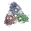
- PDB-1g2o: CRYSTAL STRUCTURE OF PURINE NUCLEOSIDE PHOSPHORYLASE FROM MYCOBAC... -
+ Open data

- Basic information
| Entry | Database: PDB / ID: 1g2o | |||||||||
|---|---|---|---|---|---|---|---|---|---|---|
| Title | CRYSTAL STRUCTURE OF PURINE NUCLEOSIDE PHOSPHORYLASE FROM MYCOBACTERIUM TUBERCULOSIS IN COMPLEX WITH A TRANSITION-STATE INHIBITOR | |||||||||
 Components Components | PURINE NUCLEOSIDE PHOSPHORYLASE | |||||||||
 Keywords Keywords | TRANSFERASE / trimer / transition-state complex | |||||||||
| Function / homology |  Function and homology information Function and homology informationdeoxyguanosine catabolic process / purine-nucleoside phosphorylase / purine-nucleoside phosphorylase activity / cytoplasm Similarity search - Function | |||||||||
| Biological species |   Mycobacterium tuberculosis (bacteria) Mycobacterium tuberculosis (bacteria) | |||||||||
| Method |  X-RAY DIFFRACTION / SYNCHROTRON / MOLECULAR REPLACEMENT / Resolution: 1.75 Å X-RAY DIFFRACTION / SYNCHROTRON / MOLECULAR REPLACEMENT / Resolution: 1.75 Å | |||||||||
 Authors Authors | Shi, W. / Basso, L.A. / Tyler, P.C. / Furneaux, R.H. / Blanchard, J.S. / Almo, S.C. / Schramm, V.L. | |||||||||
 Citation Citation | Journal: Biochemistry / Year: 2001 Title: Structures of purine nucleoside phosphorylase from Mycobacterium tuberculosis in complexes with immucillin-H and its pieces. Authors: Shi, W. / Basso, L.A. / Santos, D.S. / Tyler, P.C. / Furneaux, R.H. / Blanchard, J.S. / Almo, S.C. / Schramm, V.L. | |||||||||
| History |
|
- Structure visualization
| Structure viewer | Molecule: MolmilJmol/JSmol |
|---|
- Downloads & links
-Download
| PDBx/mmCIF format | 1g2o.cif.gz | 159.9 KB | Display | PDBx/mmCIF format |
|---|---|---|---|---|
| PDB format | pdb1g2o.ent.gz | 126.5 KB | Display | PDB format |
| PDBx/mmJSON format | 1g2o.json.gz | Tree view | PDBx/mmJSON format | |
| Others |  Other downloads Other downloads |
-Validation report
| Arichive directory | https://data.pdbj.org/pub/pdb/validation_reports/g2/1g2oftp://data.pdbj.org/pub/pdb/validation_reports/g2/1g2o | HTTPS FTP |
|---|
-Related structure data
| Related structure data |  1i80C 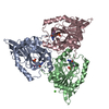 1c3xS S: Starting model for refinement C: citing same article ( |
|---|---|
| Similar structure data |


-Links
 PDBj
PDBj- Assembly
| Deposited unit | 
| ||||||||
|---|---|---|---|---|---|---|---|---|---|
| 1 |
| ||||||||
| Unit cell |
| ||||||||
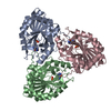
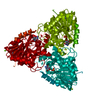
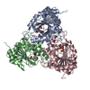
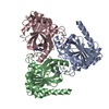
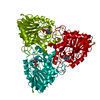
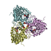
| Details | The biological assembly is a trimer in the asymmetric unit. |
-Components
| #1: Protein | Mass: 27599.457 Da / Num. of mol.: 3 Source method: isolated from a genetically manipulated source Source: (gene. exp.) Mycobacterium tuberculosis (bacteria) / Plasmid: PET-23A(+) / Species (production host): Escherichia coli / Production host: References: UniProt: P0A538, UniProt: P9WP01*PLUS, purine-nucleoside phosphorylase #2: Chemical |   Mass: 94.971 Da / Num. of mol.: 3 / Source method: obtained synthetically / Formula: PO4 Mass: 94.971 Da / Num. of mol.: 3 / Source method: obtained synthetically / Formula: PO4#3: Chemical | 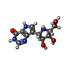  Mass: 266.253 Da / Num. of mol.: 3 / Source method: obtained synthetically / Formula: C11H14N4O4 / Comment: inhibitor*YM Mass: 266.253 Da / Num. of mol.: 3 / Source method: obtained synthetically / Formula: C11H14N4O4 / Comment: inhibitor*YM#4: Water | ChemComp-HOH / |  Mass: 18.015 Da / Num. of mol.: 485 / Source method: isolated from a natural source / Formula: H2O Mass: 18.015 Da / Num. of mol.: 485 / Source method: isolated from a natural source / Formula: H2O |
|---|
-Experimental details
-Experiment
| Experiment | Method: X-RAY DIFFRACTION / Number of used crystals: 1 |
|---|
- Sample preparation
| Crystal | Density Matthews: 2.37 Å3/Da / Density % sol: 48 % | ||||||||||||||||||||||||||||||||||||||||||
|---|---|---|---|---|---|---|---|---|---|---|---|---|---|---|---|---|---|---|---|---|---|---|---|---|---|---|---|---|---|---|---|---|---|---|---|---|---|---|---|---|---|---|---|
| Crystal grow | Temperature: 298 K / Method: vapor diffusion, hanging drop / pH: 8 Details: PEG 4000, Magnesium chloride, Tris, pH 8.0, VAPOR DIFFUSION, HANGING DROP, temperature 298K | ||||||||||||||||||||||||||||||||||||||||||
| Crystal grow | *PLUS Temperature: 18 ℃ | ||||||||||||||||||||||||||||||||||||||||||
| Components of the solutions | *PLUS
|
-Data collection
| Diffraction | Mean temperature: 100 K |
|---|---|
| Diffraction source | Source: SYNCHROTRON / Site: NSLS  / Beamline: X9B / Wavelength: 0.979 Å / Beamline: X9B / Wavelength: 0.979 Å |
| Detector | Type: ADSC QUANTUM 4 / Detector: CCD / Date: May 17, 2000 |
| Radiation | Protocol: SINGLE WAVELENGTH / Monochromatic (M) / Laue (L): M / Scattering type: x-ray |
| Radiation wavelength | Wavelength: 0.979 Å / Relative weight: 1 |
| Reflection | Resolution: 1.75→20 Å / Num. obs: 78938 / % possible obs: 99.7 % / Observed criterion σ(F): 0 / Observed criterion σ(I): 0 / Redundancy: 6.5 % / Biso Wilson estimate: 10.3 Å2 / Rsym value: 0.054 / Net I/σ(I): 35.5 |
| Reflection shell | Resolution: 1.75→1.81 Å / Redundancy: 5.5 % / Mean I/σ(I) obs: 8.3 / Num. unique all: 7823 / Rsym value: 0.217 / % possible all: 99.8 |
| Reflection | *PLUS Num. measured all: 513786 / Rmerge(I) obs: 0.054 |
| Reflection shell | *PLUS % possible obs: 99.8 % / Rmerge(I) obs: 0.217 |
- Processing
| Software |
| ||||||||||||||||||||||||||||||||||||||||
|---|---|---|---|---|---|---|---|---|---|---|---|---|---|---|---|---|---|---|---|---|---|---|---|---|---|---|---|---|---|---|---|---|---|---|---|---|---|---|---|---|---|
| Refinement | Method to determine structure: MOLECULAR REPLACEMENT Starting model: PDB ENTRY 1C3X Resolution: 1.75→20 Å / Rfactor Rfree error: 0.002 / Isotropic thermal model: Restrained / Cross valid method: THROUGHOUT / σ(F): 2 / σ(I): 1.4 / Stereochemistry target values: Engh & Huber
| ||||||||||||||||||||||||||||||||||||||||
| Solvent computation | Solvent model: FLAT MODEL / Bsol: 47.8392 Å2 / ksol: 0.392985 e/Å3 | ||||||||||||||||||||||||||||||||||||||||
| Displacement parameters | Biso mean: 14.2 Å2
| ||||||||||||||||||||||||||||||||||||||||
| Refine analyze |
| ||||||||||||||||||||||||||||||||||||||||
| Refinement step | Cycle: LAST / Resolution: 1.75→20 Å
| ||||||||||||||||||||||||||||||||||||||||
| Refine LS restraints |
| ||||||||||||||||||||||||||||||||||||||||
| LS refinement shell | Resolution: 1.75→1.86 Å / Rfactor Rfree error: 0.007 / Total num. of bins used: 6
| ||||||||||||||||||||||||||||||||||||||||
| Software | *PLUS Name: CNS / Version: 0.9 / Classification: refinement | ||||||||||||||||||||||||||||||||||||||||
| Refinement | *PLUS Lowest resolution: 20 Å / σ(F): 2 / % reflection Rfree: 10 % / Rfactor all: 0.191 / Rfactor obs: 0.189 | ||||||||||||||||||||||||||||||||||||||||
| Solvent computation | *PLUS | ||||||||||||||||||||||||||||||||||||||||
| Displacement parameters | *PLUS Biso mean: 14.2 Å2 | ||||||||||||||||||||||||||||||||||||||||
| Refine LS restraints | *PLUS
| ||||||||||||||||||||||||||||||||||||||||
| LS refinement shell | *PLUS Rfactor Rfree: 0.226 / % reflection Rfree: 10 % / Rfactor Rwork: 0.193 |
