 Movie
Movie Controller

[English] 日本語
 Yorodumi
- PDB-3scz: Combining crystallographic, thermodynamic, and molecular dynamics... -
+ Open data

- Basic information
| Entry | Database: PDB / ID: 3scz | ||||||
|---|---|---|---|---|---|---|---|
| Title | Combining crystallographic, thermodynamic, and molecular dynamics studies of Mycobacterium tuberculosis purine nucleoside phosphorylase | ||||||
 Components Components | Purine nucleoside phosphorylase | ||||||
 Keywords Keywords | TRANSFERASE / Tuberculosis / Purine nucleoside phosphorylase / Inosine / Hypoxanthine | ||||||
| Function / homology |  Function and homology information Function and homology informationdeoxyguanosine catabolic process / purine-nucleoside phosphorylase activity / purine-nucleoside phosphorylase / cytoplasm Similarity search - Function | ||||||
| Biological species |   Mycobacterium tuberculosis (bacteria) Mycobacterium tuberculosis (bacteria) | ||||||
| Method |  X-RAY DIFFRACTION / SYNCHROTRON / MOLECULAR REPLACEMENT / Resolution: 1.95 Å X-RAY DIFFRACTION / SYNCHROTRON / MOLECULAR REPLACEMENT / Resolution: 1.95 Å | ||||||
 Authors Authors | de Azevedo Jr., W.F. / Caceres, R.A. / Timmers, L.F.S.M. / Ducati, R.G. / Rosado, L.A. / Basso, L.A. / Santos, D.S. | ||||||
 Citation Citation | Journal: To be Published Title: Combining crystallographic, thermodynamic, and molecular dynamics studies of Mycobacterium tuberculosis purine nucleoside phosphorylase Authors: de Azevedo Jr., W.F. | ||||||
| History |
|
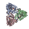
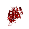
- Structure visualization
| Structure viewer | Molecule: MolmilJmol/JSmol |
|---|
- Downloads & links
-Download
| PDBx/mmCIF format | 3scz.cif.gz | 110.4 KB | Display | PDBx/mmCIF format |
|---|---|---|---|---|
| PDB format | pdb3scz.ent.gz | 86.8 KB | Display | PDB format |
| PDBx/mmJSON format | 3scz.json.gz | Tree view | PDBx/mmJSON format | |
| Others |  Other downloads Other downloads |
-Validation report
| Arichive directory | https://data.pdbj.org/pub/pdb/validation_reports/sc/3sczftp://data.pdbj.org/pub/pdb/validation_reports/sc/3scz | HTTPS FTP |
|---|
-Related structure data
| Related structure data | |
|---|---|
| Similar structure data |


-Links
 PDBj
PDBj
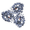
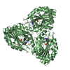
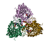
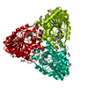
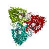
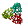
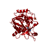
- Assembly
| Deposited unit | 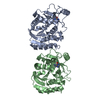
| ||||||||
|---|---|---|---|---|---|---|---|---|---|
| 1 | 
| ||||||||
| 2 | 
| ||||||||
| Unit cell |
|
-Components
| #1: Protein | Mass: 27599.457 Da / Num. of mol.: 2 Source method: isolated from a genetically manipulated source Source: (gene. exp.) Mycobacterium tuberculosis (bacteria) / Gene: punA, deoD, Rv3307, MT3406, MTV016.06 / Production host: References: UniProt: P0A538, UniProt: P9WP01*PLUS, purine-nucleoside phosphorylase #2: Chemical |   Mass: 136.111 Da / Num. of mol.: 2 / Source method: obtained synthetically / Formula: C5H4N4O Mass: 136.111 Da / Num. of mol.: 2 / Source method: obtained synthetically / Formula: C5H4N4O#3: Water | ChemComp-HOH / |  Mass: 18.015 Da / Num. of mol.: 282 / Source method: isolated from a natural source / Formula: H2O Mass: 18.015 Da / Num. of mol.: 282 / Source method: isolated from a natural source / Formula: H2O |
|---|
-Experimental details
-Experiment
| Experiment | Method: X-RAY DIFFRACTION / Number of used crystals: 1 |
|---|
- Sample preparation
| Crystal | Density Matthews: 2.01 Å3/Da / Density % sol: 38.81 % |
|---|---|
| Crystal grow | Temperature: 293 K / Method: vapor diffusion, hanging drop Details: MtPNP:INO:SO4 and MtPNP:HX were crystallized using the experimental conditions described elsewhere. PNP solution was concentrated to 25 mg/mL and co-crystallized with INO:SO4 and HX. Hanging ...Details: MtPNP:INO:SO4 and MtPNP:HX were crystallized using the experimental conditions described elsewhere. PNP solution was concentrated to 25 mg/mL and co-crystallized with INO:SO4 and HX. Hanging drops were prepared mixing 1 uL of protein solution and 1 uL of reservoir solution (100 mM Tris, pH 8.0, 25 % PEG3350, 25 mM MgCl2), VAPOR DIFFUSION, HANGING DROP, temperature 293K |
-Data collection
| Diffraction | Mean temperature: 100 K | ||||||||||||||||||||||||
|---|---|---|---|---|---|---|---|---|---|---|---|---|---|---|---|---|---|---|---|---|---|---|---|---|---|
| Diffraction source | Source: SYNCHROTRON / Site: LNLS  / Beamline: D03B-MX1 / Wavelength: 1.431 Å / Beamline: D03B-MX1 / Wavelength: 1.431 Å | ||||||||||||||||||||||||
| Detector | Type: MARMOSAIC 325 mm CCD / Detector: CCD / Date: Feb 2, 2010 | ||||||||||||||||||||||||
| Radiation | Monochromator: GRAPHITE / Protocol: SINGLE WAVELENGTH / Monochromatic (M) / Laue (L): M / Scattering type: x-ray | ||||||||||||||||||||||||
| Radiation wavelength | Wavelength: 1.431 Å / Relative weight: 1 | ||||||||||||||||||||||||
| Reflection | Resolution: 1.95→21.09 Å / Num. obs: 31352 / % possible obs: 100 % / Observed criterion σ(F): 2 / Observed criterion σ(I): 2 / Rmerge(I) obs: 0.099 / Rsym value: 0.099 | ||||||||||||||||||||||||
| Reflection shell |
|
- Processing
| Software |
| |||||||||||||||||||||||||||||||||||||||||||||||||||||||||||||||||
|---|---|---|---|---|---|---|---|---|---|---|---|---|---|---|---|---|---|---|---|---|---|---|---|---|---|---|---|---|---|---|---|---|---|---|---|---|---|---|---|---|---|---|---|---|---|---|---|---|---|---|---|---|---|---|---|---|---|---|---|---|---|---|---|---|---|---|
| Refinement | Method to determine structure: MOLECULAR REPLACEMENT / Resolution: 1.95→21.09 Å / Cor.coef. Fo:Fc: 0.946 / Cor.coef. Fo:Fc free: 0.908 / Cross valid method: THROUGHOUT / ESU R Free: 0.168 / Stereochemistry target values: MAXIMUM LIKELIHOOD / Details: HYDROGENS HAVE BEEN ADDED IN THE RIDING POSITIONS
| |||||||||||||||||||||||||||||||||||||||||||||||||||||||||||||||||
| Solvent computation | Ion probe radii: 0.8 Å / Shrinkage radii: 0.8 Å / VDW probe radii: 1.4 Å / Solvent model: MASK | |||||||||||||||||||||||||||||||||||||||||||||||||||||||||||||||||
| Displacement parameters | Biso mean: 17.071 Å2
| |||||||||||||||||||||||||||||||||||||||||||||||||||||||||||||||||
| Refinement step | Cycle: LAST / Resolution: 1.95→21.09 Å
| |||||||||||||||||||||||||||||||||||||||||||||||||||||||||||||||||
| Refine LS restraints |
| |||||||||||||||||||||||||||||||||||||||||||||||||||||||||||||||||
| LS refinement shell | Resolution: 1.95→2 Å / Total num. of bins used: 20
|
