 Movie
Movie Controller

[English] 日本語
 Yorodumi
- PDB-3iom: Crystal structure of Purine Nucleoside Phosphorylase from Mycobac... -
+ Open data

- Basic information
| Entry | Database: PDB / ID: 3iom | ||||||
|---|---|---|---|---|---|---|---|
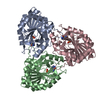
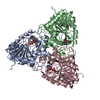
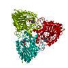
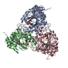
| Title | Crystal structure of Purine Nucleoside Phosphorylase from Mycobacterium tuberculosis in complex with 2'-Deoxyguanosine | ||||||
 Components Components | Purine nucleoside phosphorylase | ||||||
 Keywords Keywords | TRANSFERASE / Purine Nucleoside Phosphorylase / Mycobacterium tuberculosis / 2 -deoxyguanosine / Glycosyltransferase | ||||||
| Function / homology |  Function and homology information Function and homology informationdeoxyguanosine catabolic process / purine-nucleoside phosphorylase / purine-nucleoside phosphorylase activity / cytoplasm Similarity search - Function | ||||||
| Biological species |   Mycobacterium tuberculosis (bacteria) Mycobacterium tuberculosis (bacteria) | ||||||
| Method |  X-RAY DIFFRACTION / SYNCHROTRON / MOLECULAR REPLACEMENT / Resolution: 2.14 Å X-RAY DIFFRACTION / SYNCHROTRON / MOLECULAR REPLACEMENT / Resolution: 2.14 Å | ||||||
 Authors Authors | de Azevedo Jr., W.F. / Basso, L.A. / Santos, D.S. | ||||||
 Citation Citation | Journal: Bioorg.Med.Chem. / Year: 2010 Title: Crystallographic and docking studies of purine nucleoside phosphorylase from Mycobacterium tuberculosis. Authors: Ducati, R.G. / Basso, L.A. / Santos, D.S. / de Azevedo, W.F. | ||||||
| History |
|
- Structure visualization
| Structure viewer | Molecule: MolmilJmol/JSmol |
|---|
- Downloads & links
-Download
| PDBx/mmCIF format | 3iom.cif.gz | 111.5 KB | Display | PDBx/mmCIF format |
|---|---|---|---|---|
| PDB format | pdb3iom.ent.gz | 86.5 KB | Display | PDB format |
| PDBx/mmJSON format | 3iom.json.gz | Tree view | PDBx/mmJSON format | |
| Others |  Other downloads Other downloads |
-Validation report
| Arichive directory | https://data.pdbj.org/pub/pdb/validation_reports/io/3iomftp://data.pdbj.org/pub/pdb/validation_reports/io/3iom | HTTPS FTP |
|---|
-Related structure data
| Related structure data | 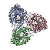 1g2oS S: Starting model for refinement |
|---|---|
| Similar structure data |


-Links
 PDBj
PDBj- Assembly
| Deposited unit | 
| ||||||||
|---|---|---|---|---|---|---|---|---|---|
| 1 | 
| ||||||||
| 2 | 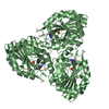
| ||||||||
| Unit cell |
| ||||||||
| Details | 2 |
-Components
| #1: Protein | Mass: 27599.457 Da / Num. of mol.: 2 Source method: isolated from a genetically manipulated source Source: (gene. exp.) Mycobacterium tuberculosis (bacteria) / Strain: Mycobacterium tuberculosisGene: deoD, MT3406, MTV016.06, punA, punA (deoD) (Rv3307) (MT3406) (MTV016.06), Rv3307 Production host: References: UniProt: P0A538, UniProt: P9WP01*PLUS, purine-nucleoside phosphorylase #2: Chemical |   Mass: 96.063 Da / Num. of mol.: 2 / Source method: obtained synthetically / Formula: SO4 Mass: 96.063 Da / Num. of mol.: 2 / Source method: obtained synthetically / Formula: SO4#3: Chemical |   Mass: 267.241 Da / Num. of mol.: 2 / Source method: obtained synthetically / Formula: C10H13N5O4 Mass: 267.241 Da / Num. of mol.: 2 / Source method: obtained synthetically / Formula: C10H13N5O4#4: Water | ChemComp-HOH / |  Mass: 18.015 Da / Num. of mol.: 209 / Source method: isolated from a natural source / Formula: H2O Mass: 18.015 Da / Num. of mol.: 209 / Source method: isolated from a natural source / Formula: H2O |
|---|
-Experimental details
-Experiment
| Experiment | Method: X-RAY DIFFRACTION / Number of used crystals: 1 |
|---|
- Sample preparation
| Crystal | Density Matthews: 1.91 Å3/Da / Density % sol: 35.55 % |
|---|---|
| Crystal grow | Temperature: 293 K / Method: vapor diffusion, hanging drop / pH: 7.6 Details: The protein was cocrystallized with a 1:0.5:1 stoichiometry of 500 mM Na2SO4 (Merck) and 3 mM 2dGuo (Fluka Biochemika), respectively, by hanging-drop vapor diffusion at 18 C. MtPNP protein ...Details: The protein was cocrystallized with a 1:0.5:1 stoichiometry of 500 mM Na2SO4 (Merck) and 3 mM 2dGuo (Fluka Biochemika), respectively, by hanging-drop vapor diffusion at 18 C. MtPNP protein (1 L of 25 mg mL-1 in 50 mM tris(hydroxymethyl)aminomethane (Tris) pH 7.6) had been previously mixed with an equal volume of the reservoir solution containing 100 mM Tris pH 8.0, 25% poly(ethylene glycol) (PEG) 3350, and 25 mM MgCl2 and hanging drops (3.5 L) were equilibrated against 400 L of the reservoir solution. , VAPOR DIFFUSION, HANGING DROP, temperature 293K |
-Data collection
| Diffraction | Mean temperature: 100 K |
|---|---|
| Diffraction source | Source: SYNCHROTRON / Site: LNLS  / Beamline: D03B-MX1 / Wavelength: 1.428 Å / Beamline: D03B-MX1 / Wavelength: 1.428 Å |
| Detector | Type: MAR CCD 165 mm / Detector: CCD / Date: Feb 2, 2009 |
| Radiation | Monochromator: GRAPHITE / Protocol: SINGLE WAVELENGTH / Monochromatic (M) / Laue (L): M / Scattering type: x-ray |
| Radiation wavelength | Wavelength: 1.428 Å / Relative weight: 1 |
| Reflection | Resolution: 2.14→28.69 Å / Num. all: 22411 / Num. obs: 22163 / % possible obs: 96.9 % / Observed criterion σ(F): 2 / Observed criterion σ(I): 2 / Redundancy: 6.8 % / Biso Wilson estimate: 21.642 Å2 / Rmerge(I) obs: 0.086 / Rsym value: 0.086 / Net I/σ(I): 5.9 |
| Reflection shell | Resolution: 2.15→2.27 Å / Redundancy: 6.4 % / Rmerge(I) obs: 0.138 / Mean I/σ(I) obs: 3.5 / Num. unique all: 625 / Rsym value: 0.138 / % possible all: 81.8 |
- Processing
| Software |
| ||||||||||||||||||||||||||||||||||||||||||||||||||||||||||||||||||||||||||||||||||||||||||
|---|---|---|---|---|---|---|---|---|---|---|---|---|---|---|---|---|---|---|---|---|---|---|---|---|---|---|---|---|---|---|---|---|---|---|---|---|---|---|---|---|---|---|---|---|---|---|---|---|---|---|---|---|---|---|---|---|---|---|---|---|---|---|---|---|---|---|---|---|---|---|---|---|---|---|---|---|---|---|---|---|---|---|---|---|---|---|---|---|---|---|---|
| Refinement | Method to determine structure: MOLECULAR REPLACEMENT Starting model: 1G2O Resolution: 2.14→28.34 Å / Cor.coef. Fo:Fc: 0.936 / Cor.coef. Fo:Fc free: 0.87 / Cross valid method: THROUGHOUT / σ(F): 2 / σ(I): 2 / ESU R: 0.352 / ESU R Free: 0.239 / Stereochemistry target values: MAXIMUM LIKELIHOOD / Details: HYDROGENS HAVE BEEN ADDED IN THE RIDING POSITIONS
| ||||||||||||||||||||||||||||||||||||||||||||||||||||||||||||||||||||||||||||||||||||||||||
| Solvent computation | Ion probe radii: 0.8 Å / Shrinkage radii: 0.8 Å / VDW probe radii: 1.2 Å / Solvent model: MASK | ||||||||||||||||||||||||||||||||||||||||||||||||||||||||||||||||||||||||||||||||||||||||||
| Displacement parameters | Biso mean: 18.181 Å2
| ||||||||||||||||||||||||||||||||||||||||||||||||||||||||||||||||||||||||||||||||||||||||||
| Refinement step | Cycle: LAST / Resolution: 2.14→28.34 Å
| ||||||||||||||||||||||||||||||||||||||||||||||||||||||||||||||||||||||||||||||||||||||||||
| Refine LS restraints |
| ||||||||||||||||||||||||||||||||||||||||||||||||||||||||||||||||||||||||||||||||||||||||||
| LS refinement shell | Resolution: 2.143→2.198 Å / Total num. of bins used: 20
|
