 Movie
Movie Controller

[English] 日本語
 Yorodumi
- PDB-3d1v: Crystal structure of human PNP complexed with 2-mercapto(3H) quin... -
+ Open data

- Basic information
| Entry | Database: PDB / ID: 3d1v | ||||||
|---|---|---|---|---|---|---|---|
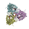
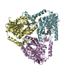
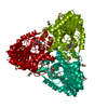
| Title | Crystal structure of human PNP complexed with 2-mercapto(3H) quinazolinone | ||||||
 Components Components | Purine nucleoside phosphorylase | ||||||
 Keywords Keywords | TRANSFERASE / PURINE NUCLEOSIDE PHOSPHORYLASE / DRUG DESIGN / SYNCHROTRON / EMPIRICAL SCORING FUNCTION / 2-MERCAPTO-4(3H) QUINAZOLINONE / Acetylation / Disease mutation / Glycosyltransferase / Polymorphism | ||||||
| Function / homology |  Function and homology information Function and homology informationnicotinamide riboside catabolic process / Defective PNP disrupts phosphorolysis of (deoxy)guanosine and (deoxy)inosine / purine-containing compound salvage / deoxyinosine catabolic process / purine nucleobase binding / nucleotide biosynthetic process / deoxyadenosine catabolic process / dAMP catabolic process / inosine catabolic process / urate biosynthetic process ...nicotinamide riboside catabolic process / Defective PNP disrupts phosphorolysis of (deoxy)guanosine and (deoxy)inosine / purine-containing compound salvage / deoxyinosine catabolic process / purine nucleobase binding / nucleotide biosynthetic process / deoxyadenosine catabolic process / dAMP catabolic process / inosine catabolic process / urate biosynthetic process / IMP catabolic process / Ribavirin ADME / guanosine phosphorylase activity / nucleoside binding / Purine salvage / allantoin metabolic process / Purine catabolism / purine-nucleoside phosphorylase / purine-nucleoside phosphorylase activity / positive regulation of alpha-beta T cell differentiation / purine ribonucleoside salvage / nucleobase-containing compound metabolic process / phosphate ion binding / positive regulation of interleukin-2 production / positive regulation of T cell proliferation / secretory granule lumen / ficolin-1-rich granule lumen / immune response / response to xenobiotic stimulus / Neutrophil degranulation / extracellular exosome / extracellular region / identical protein binding / cytoplasm / cytosol Similarity search - Function | ||||||
| Biological species |  Homo sapiens (human) Homo sapiens (human) | ||||||
| Method |  X-RAY DIFFRACTION / SYNCHROTRON / MOLECULAR REPLACEMENT / Resolution: 2.7 Å X-RAY DIFFRACTION / SYNCHROTRON / MOLECULAR REPLACEMENT / Resolution: 2.7 Å | ||||||
 Authors Authors | De Azevedo Jr., W.F. / Basso, L.A. / Santos, D.S. | ||||||
 Citation Citation | Journal: Arch.Biochem.Biophys. / Year: 2008 Title: Structural studies of human purine nucleoside phosphorylase: towards a new specific empirical scoring function Authors: Timmers, L.F. / Caceres, R.A. / Vivan, A.L. / Gava, L.M. / Dias, R. / Ducati, R.G. / Basso, L.A. / Santos, D.S. / de Azevedo, W.F. #1: Journal: Biochem.Biophys.Res.Commun. / Year: 2003Title: Crystal Structure of Human Purine Nucleoside Phosphorylase at 2.3A Resolution Authors: De Azevedo Jr., W.F. / Canduri, F. / Dos Santos, D.M. / Silva, R.G. / De Oliveira, J.S. / De Carvalho, L.P.S. / Basso, L.A. / Mendes, M.A. / Palma, M.S. / Santos, D.S. | ||||||
| History |
|
- Structure visualization
| Structure viewer | Molecule: MolmilJmol/JSmol |
|---|
- Downloads & links
-Download
| PDBx/mmCIF format | 3d1v.cif.gz | 75.2 KB | Display | PDBx/mmCIF format |
|---|---|---|---|---|
| PDB format | pdb3d1v.ent.gz | 55 KB | Display | PDB format |
| PDBx/mmJSON format | 3d1v.json.gz | Tree view | PDBx/mmJSON format | |
| Others |  Other downloads Other downloads |
-Validation report
| Arichive directory | https://data.pdbj.org/pub/pdb/validation_reports/d1/3d1vftp://data.pdbj.org/pub/pdb/validation_reports/d1/3d1v | HTTPS FTP |
|---|
-Related structure data
| Related structure data |  1m73S S: Starting model for refinement |
|---|---|
| Similar structure data |


-Links
 PDBj
PDBj
- Assembly
| Deposited unit | 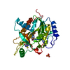
| ||||||||
|---|---|---|---|---|---|---|---|---|---|
| 1 | 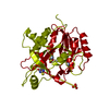
| ||||||||
| Unit cell |
|
-Components
| #1: Protein | Mass: 32184.877 Da / Num. of mol.: 1 Source method: isolated from a genetically manipulated source Source: (gene. exp.) Homo sapiens (human) / Gene: NP, PNP / Production host:  References: UniProt: P00491, purine-nucleoside phosphorylase | ||||
|---|---|---|---|---|---|
| #2: Chemical |   Mass: 96.063 Da / Num. of mol.: 3 / Source method: obtained synthetically / Formula: SO4 Mass: 96.063 Da / Num. of mol.: 3 / Source method: obtained synthetically / Formula: SO4#3: Chemical | ChemComp-D1V / |   Mass: 178.211 Da / Num. of mol.: 1 / Source method: obtained synthetically / Formula: C8H6N2OS Mass: 178.211 Da / Num. of mol.: 1 / Source method: obtained synthetically / Formula: C8H6N2OS#4: Water | ChemComp-HOH / |  Mass: 18.015 Da / Num. of mol.: 69 / Source method: isolated from a natural source / Formula: H2O Mass: 18.015 Da / Num. of mol.: 69 / Source method: isolated from a natural source / Formula: H2O |
-Experimental details
-Experiment
| Experiment | Method: X-RAY DIFFRACTION / Number of used crystals: 1 |
|---|
- Sample preparation
| Crystal | Density Matthews: 4.92 Å3/Da / Density % sol: 75 % |
|---|---|
| Crystal grow | Method: vapor diffusion, hanging drop / pH: 5.3 Details: 0.05M CITRATE BUFFER, 19%AMMONIUM SULPHATE, PH 5.30, VAPOR DIFFUSION, HANGING DROP |
-Data collection
| Diffraction | Mean temperature: 104 K |
|---|---|
| Diffraction source | Source: SYNCHROTRON / Site: LNLS  / Beamline: D03B-MX1 / Wavelength: 1.431 / Beamline: D03B-MX1 / Wavelength: 1.431 |
| Detector | Type: MARRESEARCH / Detector: CCD / Date: Apr 16, 2005 |
| Radiation | Monochromator: GRAPHITE / Protocol: SINGLE WAVELENGTH / Monochromatic (M) / Laue (L): M / Scattering type: x-ray |
| Radiation wavelength | Wavelength: 1.431 Å / Relative weight: 1 |
| Reflection | Resolution: 2.7→56.531 Å / Num. obs: 16763 / % possible obs: 90.1 % / Observed criterion σ(I): 2 / Redundancy: 5.5 % / Rsym value: 0.099 / Net I/σ(I): 6.6 |
| Reflection shell | Resolution: 2.7→2.84 Å / Redundancy: 5.3 % / Mean I/σ(I) obs: 1.9 / Rsym value: 0.375 / % possible all: 93.4 |
- Processing
| Software |
| ||||||||||||||||||||
|---|---|---|---|---|---|---|---|---|---|---|---|---|---|---|---|---|---|---|---|---|---|
| Refinement | Method to determine structure: MOLECULAR REPLACEMENT Starting model: PDB entry 1M73 Resolution: 2.7→42.18 Å / Cor.coef. Fo:Fc: 0.921 / Cor.coef. Fo:Fc free: 0.879 / SU B: 9.454 / SU ML: 0.201 / Cross valid method: THROUGHOUT / ESU R: 0.382 / ESU R Free: 0.299 / Stereochemistry target values: MAXIMUM LIKELIHOOD Details: HYDROGENS HAVE BEEN ADDED IN THE RIDING POSITIONS. The distance of the C-N bond between PRO146 and LEU147 on chain A is 1.91 Angstrom. The distance of the C-N bond between ALA196 and GLY197 ...Details: HYDROGENS HAVE BEEN ADDED IN THE RIDING POSITIONS. The distance of the C-N bond between PRO146 and LEU147 on chain A is 1.91 Angstrom. The distance of the C-N bond between ALA196 and GLY197 on chain A is 1.63 Angstrom. The distance of the C-N bond between PRO198 and SER199 on chain A is 1.68 Angstrom. The distance of the C-N bond between SER220 and THR221 on chain A is 1.20 Angstrom. The distance of the C-N bond between PRO223 and GLU224 on chain A is 1.18 Angstrom.
| ||||||||||||||||||||
| Solvent computation | Ion probe radii: 0.8 Å / Shrinkage radii: 0.8 Å / VDW probe radii: 1.4 Å / Solvent model: MASK | ||||||||||||||||||||
| Displacement parameters | Biso mean: 44.179 Å2
| ||||||||||||||||||||
| Refinement step | Cycle: LAST / Resolution: 2.7→42.18 Å
| ||||||||||||||||||||
| LS refinement shell | Resolution: 2.7→2.77 Å / Total num. of bins used: 20
|
