 Movie
Movie Controller


+ Open data

- Basic information
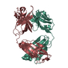
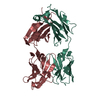
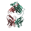
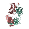
| Entry | Database: PDB / ID: 1dqq | ||||||
|---|---|---|---|---|---|---|---|
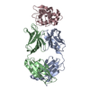
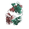
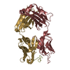
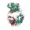
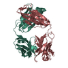
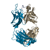
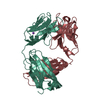
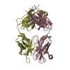
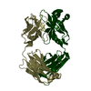
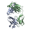
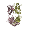
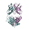
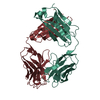
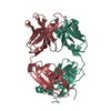
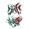
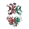
| Title | CRYSTAL STRUCTURE OF ANTI-LYSOZYME ANTIBODY HYHEL-63 | ||||||
 Components Components |
| ||||||
 Keywords Keywords | IMMUNE SYSTEM / anti-lysozyme antibody / HyHEL-63 / hen egg white lysozyme | ||||||
| Function / homology |  Function and homology information Function and homology informationalpha-beta T cell receptor complex / immunoglobulin receptor binding / IgG immunoglobulin complex / B cell differentiation / adaptive immune response / extracellular region / plasma membrane Similarity search - Function | ||||||
| Biological species |  | ||||||
| Method |  X-RAY DIFFRACTION / Resolution: 1.8 Å X-RAY DIFFRACTION / Resolution: 1.8 Å | ||||||
 Authors Authors | Li, H. / Mariuzza, R.A. | ||||||
 Citation Citation | Journal: Biochemistry / Year: 2000 Title: Three-dimensional structures of the free and antigen-bound Fab from monoclonal antilysozyme antibody HyHEL-63(,). Authors: Li, Y. / Li, H. / Smith-Gill, S.J. / Mariuzza, R.A. | ||||||
| History |
|
- Structure visualization
| Structure viewer | Molecule: MolmilJmol/JSmol |
|---|
- Downloads & links
-Download
| PDBx/mmCIF format | 1dqq.cif.gz | 193.4 KB | Display | PDBx/mmCIF format |
|---|---|---|---|---|
| PDB format | pdb1dqq.ent.gz | 153.3 KB | Display | PDB format |
| PDBx/mmJSON format | 1dqq.json.gz | Tree view | PDBx/mmJSON format | |
| Others |  Other downloads Other downloads |
-Validation report
| Arichive directory | https://data.pdbj.org/pub/pdb/validation_reports/dq/1dqqftp://data.pdbj.org/pub/pdb/validation_reports/dq/1dqq | HTTPS FTP |
|---|
-Related structure data


-Links
 PDBj
PDBj


- Assembly
| Deposited unit | 
| ||||||||
|---|---|---|---|---|---|---|---|---|---|
| 1 | 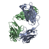
| ||||||||
| 2 | 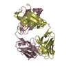
| ||||||||
| Unit cell |
| ||||||||
| Details | The biological assembly is a heterodimer composed of heavy and light chains. |
-Components
| #1: Antibody | Mass: 23583.869 Da / Num. of mol.: 2 / Fragment: FAB FRAGMENT Source method: isolated from a genetically manipulated source Source: (gene. exp.)  #2: Antibody | Mass: 22556.023 Da / Num. of mol.: 2 / Fragment: FAB FRAGMENT Source method: isolated from a genetically manipulated source Source: (gene. exp.) #3: Water | ChemComp-HOH / |  Mass: 18.015 Da / Num. of mol.: 858 / Source method: isolated from a natural source / Formula: H2O Mass: 18.015 Da / Num. of mol.: 858 / Source method: isolated from a natural source / Formula: H2OHas protein modification | Y | |
|---|
-Experimental details
-Experiment
| Experiment | Method: X-RAY DIFFRACTION / Number of used crystals: 1 |
|---|
- Sample preparation
| Crystal | Density Matthews: 2.37 Å3/Da / Density % sol: 48.09 % | ||||||||||||||||||||||||||||||
|---|---|---|---|---|---|---|---|---|---|---|---|---|---|---|---|---|---|---|---|---|---|---|---|---|---|---|---|---|---|---|---|
| Crystal grow | Temperature: 298 K / Method: vapor diffusion, hanging drop / pH: 7.5 Details: PEG 8000, KH2PO4, Tris-HCl, pH 7.5, VAPOR DIFFUSION, HANGING DROP, temperature 298K | ||||||||||||||||||||||||||||||
| Crystal grow | *PLUS Details: drop consists of equal volume of protein and reservoir solutions | ||||||||||||||||||||||||||||||
| Components of the solutions | *PLUS
|
-Data collection
| Diffraction | Mean temperature: 100 K |
|---|---|
| Diffraction source | Source: ROTATING ANODE / Type: RIGAKU RU200 / Wavelength: 1.5418 |
| Detector | Type: MACSCIENCE / Detector: IMAGE PLATE / Date: Jun 6, 1999 |
| Radiation | Protocol: SINGLE WAVELENGTH / Monochromatic (M) / Laue (L): M / Scattering type: x-ray |
| Radiation wavelength | Wavelength: 1.5418 Å / Relative weight: 1 |
| Reflection | Resolution: 1.8→100 Å / Num. all: 68027 / Num. obs: 145275 / % possible obs: 86.5 % / Observed criterion σ(F): 2 / Observed criterion σ(I): 2 / Redundancy: 2.1 % / Biso Wilson estimate: 20.4 Å2 / Rmerge(I) obs: 0.049 / Net I/σ(I): 16 |
| Reflection shell | Resolution: 1.8→1.9 Å / Redundancy: 1.5 % / Rmerge(I) obs: 0.02 / % possible all: 54.5 |
| Reflection | *PLUS Num. obs: 68027 / Num. measured all: 145275 |
| Reflection shell | *PLUS % possible obs: 54.5 % / Rmerge(I) obs: 0.315 / Mean I/σ(I) obs: 2 |
- Processing
| Software |
| |||||||||||||||||||||||||
|---|---|---|---|---|---|---|---|---|---|---|---|---|---|---|---|---|---|---|---|---|---|---|---|---|---|---|
| Refinement | Resolution: 1.8→100 Å / σ(F): 0 / σ(I): 0 / Stereochemistry target values: Engh & Huber
| |||||||||||||||||||||||||
| Refinement step | Cycle: LAST / Resolution: 1.8→100 Å
| |||||||||||||||||||||||||
| Refine LS restraints |
| |||||||||||||||||||||||||
| Software | *PLUS Name: 'CNS' / Classification: refinement | |||||||||||||||||||||||||
| Refinement | *PLUS | |||||||||||||||||||||||||
| Solvent computation | *PLUS | |||||||||||||||||||||||||
| Displacement parameters | *PLUS Biso mean: 26.7 Å2 | |||||||||||||||||||||||||
| Refine LS restraints | *PLUS
|
