 Movie
Movie Controller

[English] 日本語
 Yorodumi
- PDB-4yxl: Crystal structure of Syrian hamster prion protein complexed with ... -
+ Open data

- Basic information
| Entry | Database: PDB / ID: 4yxl | ||||||
|---|---|---|---|---|---|---|---|
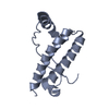
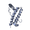
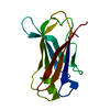
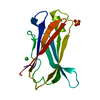
| Title | Crystal structure of Syrian hamster prion protein complexed with POM1 FAB | ||||||
 Components Components |
| ||||||
 Keywords Keywords | IMMUNE SYSTEM / prion / antibody / Immune system complex | ||||||
| Function / homology |  Function and homology information Function and homology informationaspartic-type endopeptidase inhibitor activity / regulation of calcium ion import across plasma membrane / glycosaminoglycan binding / positive regulation of glutamate receptor signaling pathway / negative regulation of interleukin-17 production / cupric ion binding / regulation of potassium ion transmembrane transport / type 5 metabotropic glutamate receptor binding / negative regulation of dendritic spine maintenance / negative regulation of calcineurin-NFAT signaling cascade ...aspartic-type endopeptidase inhibitor activity / regulation of calcium ion import across plasma membrane / glycosaminoglycan binding / positive regulation of glutamate receptor signaling pathway / negative regulation of interleukin-17 production / cupric ion binding / regulation of potassium ion transmembrane transport / type 5 metabotropic glutamate receptor binding / negative regulation of dendritic spine maintenance / negative regulation of calcineurin-NFAT signaling cascade / negative regulation of interleukin-2 production / negative regulation of activated T cell proliferation / negative regulation of amyloid-beta formation / negative regulation of type II interferon production / cuprous ion binding / positive regulation of protein targeting to membrane / negative regulation of T cell receptor signaling pathway / side of membrane / neuron projection maintenance / inclusion body / positive regulation of calcium-mediated signaling / molecular function activator activity / cellular response to copper ion / positive regulation of protein localization to plasma membrane / molecular condensate scaffold activity / protein homooligomerization / protein destabilization / cellular response to xenobiotic stimulus / cellular response to amyloid-beta / terminal bouton / positive regulation of neuron apoptotic process / amyloid-beta binding / presynapse / signaling receptor activity / protease binding / response to oxidative stress / nuclear membrane / microtubule binding / amyloid fibril formation / learning or memory / regulation of cell cycle / intracellular signal transduction / membrane raft / copper ion binding / dendrite / negative regulation of apoptotic process / protein-containing complex binding / cell surface / negative regulation of transcription by RNA polymerase II / Golgi apparatus / endoplasmic reticulum / identical protein binding / plasma membrane / cytosol Similarity search - Function | ||||||
| Biological species |  Mesocricetus auratus (golden hamster) Mesocricetus auratus (golden hamster) | ||||||
| Method |  X-RAY DIFFRACTION / SYNCHROTRON / MOLECULAR REPLACEMENT / Resolution: 2.604 Å X-RAY DIFFRACTION / SYNCHROTRON / MOLECULAR REPLACEMENT / Resolution: 2.604 Å | ||||||
 Authors Authors | Baral, P.K. / Swayampakula, M. / James, M.N.G. | ||||||
| Funding support |  Canada, 1items Canada, 1items
| ||||||
 Citation Citation | Journal: J.Struct.Biol. / Year: 2015 Title: X-ray structural and molecular dynamical studies of the globular domains of cow, deer, elk and Syrian hamster prion proteins. Authors: Baral, P.K. / Swayampakula, M. / Aguzzi, A. / James, M.N. | ||||||
| History |
|
- Structure visualization
| Structure viewer | Molecule: MolmilJmol/JSmol |
|---|
- Downloads & links
-Download
| PDBx/mmCIF format | 4yxl.cif.gz | 121 KB | Display | PDBx/mmCIF format |
|---|---|---|---|---|
| PDB format | pdb4yxl.ent.gz | 91.4 KB | Display | PDB format |
| PDBx/mmJSON format | 4yxl.json.gz | Tree view | PDBx/mmJSON format | |
| Others |  Other downloads Other downloads |
-Validation report
| Arichive directory | https://data.pdbj.org/pub/pdb/validation_reports/yx/4yxlftp://data.pdbj.org/pub/pdb/validation_reports/yx/4yxl | HTTPS FTP |
|---|
-Related structure data
| Related structure data |  4yx2C  4yxhC  4yxkC  4dgiS  4h88S C: citing same article ( S: Starting model for refinement |
|---|---|
| Similar structure data |


-Links
 PDBj
PDBj


- Assembly
| Deposited unit | 
| ||||||||
|---|---|---|---|---|---|---|---|---|---|
| 1 | 
| ||||||||
| 2 | 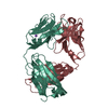
| ||||||||
| Unit cell |
|
-Components
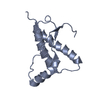
| #1: Protein | Mass: 18896.012 Da / Num. of mol.: 1 Source method: isolated from a genetically manipulated source Source: (gene. exp.) Mesocricetus auratus (golden hamster) / Gene: PRNP, PRP / Production host:  |
|---|---|
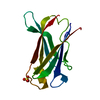
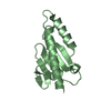
| #2: Antibody | Mass: 23367.053 Da / Num. of mol.: 1 Source method: isolated from a genetically manipulated source Source: (gene. exp.) |
| #3: Antibody | Mass: 23509.701 Da / Num. of mol.: 1 Source method: isolated from a genetically manipulated source Source: (gene. exp.) |
| #4: Chemical | ChemComp-NA /   Mass: 22.990 Da / Num. of mol.: 1 / Source method: obtained synthetically / Formula: Na Mass: 22.990 Da / Num. of mol.: 1 / Source method: obtained synthetically / Formula: Na |
| #5: Water | ChemComp-HOH /  Mass: 18.015 Da / Num. of mol.: 76 / Source method: isolated from a natural source / Formula: H2O Mass: 18.015 Da / Num. of mol.: 76 / Source method: isolated from a natural source / Formula: H2O |
| Has protein modification | Y |
-Experimental details
-Experiment
| Experiment | Method: X-RAY DIFFRACTION |
|---|
- Sample preparation
| Crystal | Density Matthews: 2.82 Å3/Da / Density % sol: 56.36 % |
|---|---|
| Crystal grow | Temperature: 295 K / Method: vapor diffusion, sitting drop Details: 25% PEG3350, 0.1 M BIS-TRIS, 0.2 M lithium sulphate |
-Data collection
| Diffraction | Mean temperature: 100 K |
|---|---|
| Diffraction source | Source: SYNCHROTRON / Site: SSRL  / Beamline: BL9-1 / Wavelength: 0.97947 Å / Beamline: BL9-1 / Wavelength: 0.97947 Å |
| Detector | Type: MARMOSAIC 325 mm CCD / Detector: CCD / Date: May 30, 2011 |
| Radiation | Protocol: SINGLE WAVELENGTH / Monochromatic (M) / Laue (L): M / Scattering type: x-ray |
| Radiation wavelength | Wavelength: 0.97947 Å / Relative weight: 1 |
| Reflection | Resolution: 2.6→50 Å / Num. obs: 19709 / % possible obs: 97.4 % / Redundancy: 4 % / Rsym value: 0.08 / Net I/σ(I): 15.46 |
| Reflection shell | Resolution: 2.6→2.69 Å / Redundancy: 3.7 % / Mean I/σ(I) obs: 1.44 / % possible all: 87.5 |
- Processing
| Software |
| ||||||||||||||||||||||||||||||||||||||||||||||||||||||||
|---|---|---|---|---|---|---|---|---|---|---|---|---|---|---|---|---|---|---|---|---|---|---|---|---|---|---|---|---|---|---|---|---|---|---|---|---|---|---|---|---|---|---|---|---|---|---|---|---|---|---|---|---|---|---|---|---|---|
| Refinement | Method to determine structure: MOLECULAR REPLACEMENT Starting model: 4H88, 4DGI Resolution: 2.604→35.241 Å / SU ML: 0.32 / Cross valid method: FREE R-VALUE / σ(F): 1.35 / Phase error: 38.23 / Stereochemistry target values: ML
| ||||||||||||||||||||||||||||||||||||||||||||||||||||||||
| Solvent computation | Shrinkage radii: 0.9 Å / VDW probe radii: 1.11 Å / Solvent model: FLAT BULK SOLVENT MODEL | ||||||||||||||||||||||||||||||||||||||||||||||||||||||||
| Refinement step | Cycle: LAST / Resolution: 2.604→35.241 Å
| ||||||||||||||||||||||||||||||||||||||||||||||||||||||||
| Refine LS restraints |
| ||||||||||||||||||||||||||||||||||||||||||||||||||||||||
| LS refinement shell |
|
