 Movie
Movie Controller


+ Open data

- Basic information
| Entry | Database: PDB / ID: 1d1s | ||||||
|---|---|---|---|---|---|---|---|
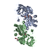
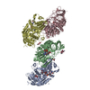
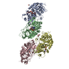
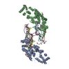
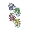
| Title | WILD-TYPE HUMAN SIGMA (CLASS IV) ALCOHOL DEHYDROGENASE | ||||||
 Components Components | ALCOHOL DEHYDROGENASE CLASS IV SIGMA CHAIN | ||||||
 Keywords Keywords | OXIDOREDUCTASE / ROSSMAN OR DINUCLEOTIDE FOLD | ||||||
| Function / homology |  Function and homology information Function and homology informationomega-hydroxydecanoate dehydrogenase / omega-hydroxydecanoate dehydrogenase activity / ethanol binding / aldehyde oxidase activity / all-trans-retinol dehydrogenase (NAD+) / fatty acid omega-oxidation / receptor antagonist activity / Ethanol oxidation / alcohol dehydrogenase (NAD+) activity / all-trans-retinol dehydrogenase (NAD+) activity ...omega-hydroxydecanoate dehydrogenase / omega-hydroxydecanoate dehydrogenase activity / ethanol binding / aldehyde oxidase activity / all-trans-retinol dehydrogenase (NAD+) / fatty acid omega-oxidation / receptor antagonist activity / Ethanol oxidation / alcohol dehydrogenase (NAD+) activity / all-trans-retinol dehydrogenase (NAD+) activity / alcohol dehydrogenase / retinoic acid metabolic process / retinol metabolic process / retinol binding / retinoid metabolic process / response to bacterium / response to ethanol / zinc ion binding / plasma membrane / cytosol Similarity search - Function | ||||||
| Biological species |  Homo sapiens (human) Homo sapiens (human) | ||||||
| Method |  X-RAY DIFFRACTION / Resolution: 2.5 Å X-RAY DIFFRACTION / Resolution: 2.5 Å | ||||||
 Authors Authors | Xie, P.T. / Hurley, T.D. | ||||||
 Citation Citation | Journal: Protein Sci. / Year: 1999 Title: Methionine-141 directly influences the binding of 4-methylpyrazole in human sigma sigma alcohol dehydrogenase. Authors: Xie, P.T. / Hurley, T.D. #1: Journal: J.Biol.Chem. / Year: 1997Title: X-ray structure of human class IV sigma-sigma alcohol dehydrogenase Authors: Xie, P.T. / Parsons, S.H. / Speckhard, D.C. / Bosron, W.F. / Hurley, T.D. | ||||||
| History |
|
- Structure visualization
| Structure viewer | Molecule: MolmilJmol/JSmol |
|---|
- Downloads & links
-Download
| PDBx/mmCIF format | 1d1s.cif.gz | 302.8 KB | Display | PDBx/mmCIF format |
|---|---|---|---|---|
| PDB format | pdb1d1s.ent.gz | 245.6 KB | Display | PDB format |
| PDBx/mmJSON format | 1d1s.json.gz | Tree view | PDBx/mmJSON format | |
| Others |  Other downloads Other downloads |
-Validation report
| Arichive directory | https://data.pdbj.org/pub/pdb/validation_reports/d1/1d1sftp://data.pdbj.org/pub/pdb/validation_reports/d1/1d1s | HTTPS FTP |
|---|
-Related structure data

-Links
 PDBj
PDBj


- Assembly
| Deposited unit | 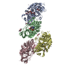
| ||||||||
|---|---|---|---|---|---|---|---|---|---|
| 1 |
| ||||||||
| 2 | 
| ||||||||
| 3 | 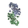
| ||||||||
| 4 | 
| ||||||||
| Unit cell |
|
-Components
-Protein , 1 types, 4 molecules ABCD
| #1: Protein | Mass: 39924.352 Da / Num. of mol.: 4 Source method: isolated from a genetically manipulated source Source: (gene. exp.) Homo sapiens (human) / Production host:  |
|---|
-Non-polymers , 5 types, 449 molecules 


| #2: Chemical | ChemComp-ZN /  Mass: 65.409 Da / Num. of mol.: 19 / Source method: obtained synthetically / Formula: Zn Mass: 65.409 Da / Num. of mol.: 19 / Source method: obtained synthetically / Formula: Zn#3: Chemical | ChemComp-ACT /  Mass: 59.044 Da / Num. of mol.: 13 / Source method: obtained synthetically / Formula: C2H3O2 Mass: 59.044 Da / Num. of mol.: 13 / Source method: obtained synthetically / Formula: C2H3O2#4: Chemical | ChemComp-CAC /  Mass: 136.989 Da / Num. of mol.: 5 / Source method: obtained synthetically / Formula: C2H6AsO2 Mass: 136.989 Da / Num. of mol.: 5 / Source method: obtained synthetically / Formula: C2H6AsO2#5: Chemical | ChemComp-NAD /  Mass: 663.425 Da / Num. of mol.: 4 / Source method: obtained synthetically / Formula: C21H27N7O14P2 / Comment: NAD*YM Mass: 663.425 Da / Num. of mol.: 4 / Source method: obtained synthetically / Formula: C21H27N7O14P2 / Comment: NAD*YM#6: Water | ChemComp-HOH / | Mass: 18.015 Da / Num. of mol.: 408 / Source method: isolated from a natural source / Formula: H2O |
|---|
-Experimental details
-Experiment
| Experiment | Method: X-RAY DIFFRACTION / Number of used crystals: 1 |
|---|
- Sample preparation
| Crystal | Density Matthews: 2.93 Å3/Da / Density % sol: 58.09 % | ||||||||||||||||||||||||||||||
|---|---|---|---|---|---|---|---|---|---|---|---|---|---|---|---|---|---|---|---|---|---|---|---|---|---|---|---|---|---|---|---|
| Crystal grow | Temperature: 277 K / Method: vapor diffusion, sitting drop / pH: 6.5 Details: 100 MM CACODYLATE, 100 MM ZINC ACETATE, 7.5 MM NAD+, 18% POLYETHYLENE GLYCOL 6000, 8 MG/ML ENZYME, pH 6.5, VAPOR DIFFUSION, SITTING DROP, temperature 277K | ||||||||||||||||||||||||||||||
| Crystal grow | *PLUS Temperature: 4 ℃ / Method: unknown | ||||||||||||||||||||||||||||||
| Components of the solutions | *PLUS
|
-Data collection
| Diffraction | Mean temperature: 113 K |
|---|---|
| Diffraction source | Source: ROTATING ANODE / Type: RIGAKU RU200 / Wavelength: 1.5418 |
| Detector | Type: RIGAKU RAXIS IIC / Detector: IMAGE PLATE / Date: Aug 10, 1997 |
| Radiation | Protocol: SINGLE WAVELENGTH / Monochromatic (M) / Laue (L): M / Scattering type: x-ray |
| Radiation wavelength | Wavelength: 1.5418 Å / Relative weight: 1 |
| Reflection | Resolution: 2.5→50 Å / Num. all: 64199 / Num. obs: 58678 / % possible obs: 91.4 % / Observed criterion σ(F): 0 / Observed criterion σ(I): 0 / Redundancy: 4.33 % / Biso Wilson estimate: 42.2 Å2 / Rmerge(I) obs: 0.085 / Net I/σ(I): 17.1 |
| Reflection shell | Resolution: 2.5→2.6 Å / Redundancy: 2.5 % / Rmerge(I) obs: 0.368 / % possible all: 76 |
| Reflection | *PLUS Num. measured all: 254379 |
| Reflection shell | *PLUS % possible obs: 76 % / Mean I/σ(I) obs: 2.6 |
- Processing
| Software |
| ||||||||||||||||||||||||||||||||||||||||||||||||||||||||||||
|---|---|---|---|---|---|---|---|---|---|---|---|---|---|---|---|---|---|---|---|---|---|---|---|---|---|---|---|---|---|---|---|---|---|---|---|---|---|---|---|---|---|---|---|---|---|---|---|---|---|---|---|---|---|---|---|---|---|---|---|---|---|
| Refinement | Resolution: 2.5→50 Å / σ(F): 0.5 / σ(I): 0.5 / Stereochemistry target values: ENGH AND HUBER
| ||||||||||||||||||||||||||||||||||||||||||||||||||||||||||||
| Refinement step | Cycle: LAST / Resolution: 2.5→50 Å
| ||||||||||||||||||||||||||||||||||||||||||||||||||||||||||||
| Refine LS restraints |
| ||||||||||||||||||||||||||||||||||||||||||||||||||||||||||||
| Software | *PLUS Name: X-PLOR / Version: 3.851 / Classification: refinement | ||||||||||||||||||||||||||||||||||||||||||||||||||||||||||||
| Refine LS restraints | *PLUS
|
