 Movie
Movie Controller

[English] 日本語
 Yorodumi
- PDB-3kt3: Crystal structure of S. cerevisiae tryptophanyl-tRNA synthetase i... -
+ Open data

- Basic information
| Entry | Database: PDB / ID: 3kt3 | ||||||
|---|---|---|---|---|---|---|---|
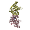
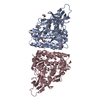
| Title | Crystal structure of S. cerevisiae tryptophanyl-tRNA synthetase in complex with TrpAMP | ||||||
 Components Components | Tryptophanyl-tRNA synthetase, cytoplasmic | ||||||
 Keywords Keywords | LIGASE / tryptophanyl-tRNA synthetase / S. cerevisiae / sulfate ion / amino acid activation / catalytic mechanism / Aminoacyl-tRNA synthetase / ATP-binding / Cytoplasm / Nucleotide-binding / Protein biosynthesis | ||||||
| Function / homology |  Function and homology information Function and homology informationtryptophan-tRNA ligase / tryptophanyl-tRNA aminoacylation / tryptophan-tRNA ligase activity / sequence-specific mRNA binding / ATP binding / cytoplasm Similarity search - Function | ||||||
| Biological species |  | ||||||
| Method |  X-RAY DIFFRACTION / SYNCHROTRON / MOLECULAR REPLACEMENT / Resolution: 2.6 Å X-RAY DIFFRACTION / SYNCHROTRON / MOLECULAR REPLACEMENT / Resolution: 2.6 Å | ||||||
 Authors Authors | Zhou, M. / Dong, X. / Zhong, C. / Shen, N. / Ding, J. | ||||||
 Citation Citation | Journal: Nucleic Acids Res. / Year: 2010 Title: Crystal structures of Saccharomyces cerevisiae tryptophanyl-tRNA synthetase: new insights into the mechanism of tryptophan activation and implications for anti-fungal drug design Authors: Zhou, M. / Dong, X. / Shen, N. / Zhong, C. / Ding, J. | ||||||
| History |
|
- Structure visualization
| Structure viewer | Molecule: MolmilJmol/JSmol |
|---|
- Downloads & links
-Download
| PDBx/mmCIF format | 3kt3.cif.gz | 624.4 KB | Display | PDBx/mmCIF format |
|---|---|---|---|---|
| PDB format | pdb3kt3.ent.gz | 519 KB | Display | PDB format |
| PDBx/mmJSON format | 3kt3.json.gz | Tree view | PDBx/mmJSON format | |
| Others |  Other downloads Other downloads |
-Validation report
| Arichive directory | https://data.pdbj.org/pub/pdb/validation_reports/kt/3kt3ftp://data.pdbj.org/pub/pdb/validation_reports/kt/3kt3 | HTTPS FTP |
|---|
-Related structure data
| Related structure data | 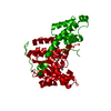 3kt0SC 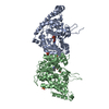 3kt6C  3kt8C S: Starting model for refinement C: citing same article ( |
|---|---|
| Similar structure data |


-Links
 PDBj
PDBj
- Assembly
| Deposited unit | 
| ||||||||
|---|---|---|---|---|---|---|---|---|---|
| 1 | 
| ||||||||
| 2 | 
| ||||||||
| Unit cell |
|
-Components
| #1: Protein | Mass: 50248.094 Da / Num. of mol.: 4 Source method: isolated from a genetically manipulated source Source: (gene. exp.) Gene: trpS / Plasmid: pET3E / Production host:  #2: Chemical | ChemComp-TYM /   Mass: 533.431 Da / Num. of mol.: 4 / Source method: obtained synthetically / Formula: C21H24N7O8P Mass: 533.431 Da / Num. of mol.: 4 / Source method: obtained synthetically / Formula: C21H24N7O8P#3: Chemical | ChemComp-SO4 /   Mass: 96.063 Da / Num. of mol.: 13 / Source method: obtained synthetically / Formula: SO4 Mass: 96.063 Da / Num. of mol.: 13 / Source method: obtained synthetically / Formula: SO4#4: Water | ChemComp-HOH / |  Mass: 18.015 Da / Num. of mol.: 178 / Source method: isolated from a natural source / Formula: H2O Mass: 18.015 Da / Num. of mol.: 178 / Source method: isolated from a natural source / Formula: H2O |
|---|
-Experimental details
-Experiment
| Experiment | Method: X-RAY DIFFRACTION / Number of used crystals: 1 |
|---|
- Sample preparation
| Crystal | Density Matthews: 5.056929 Å3/Da / Density % sol: 75.676941 % / Mosaicity: 0.608 ° |
|---|---|
| Crystal grow | Temperature: 277 K / Method: vapor diffusion, hanging drop / pH: 5.9 Details: 0.1M HEPES, 2.2M (NH4)2SO4, pH 5.9, VAPOR DIFFUSION, HANGING DROP, temperature 277K |
-Data collection
| Diffraction | Mean temperature: 100 K | ||||||||||||||||||||||||||||||||||||||||||||||||||||||||||||||||||
|---|---|---|---|---|---|---|---|---|---|---|---|---|---|---|---|---|---|---|---|---|---|---|---|---|---|---|---|---|---|---|---|---|---|---|---|---|---|---|---|---|---|---|---|---|---|---|---|---|---|---|---|---|---|---|---|---|---|---|---|---|---|---|---|---|---|---|---|
| Diffraction source | Source: SYNCHROTRON / Site: Photon Factory  / Beamline: AR-NW12A / Wavelength: 1 Å / Beamline: AR-NW12A / Wavelength: 1 Å | ||||||||||||||||||||||||||||||||||||||||||||||||||||||||||||||||||
| Detector | Type: ADSC QUANTUM 210r / Detector: CCD / Date: Nov 23, 2008 | ||||||||||||||||||||||||||||||||||||||||||||||||||||||||||||||||||
| Radiation | Protocol: SINGLE WAVELENGTH / Monochromatic (M) / Laue (L): M / Scattering type: x-ray | ||||||||||||||||||||||||||||||||||||||||||||||||||||||||||||||||||
| Radiation wavelength | Wavelength: 1 Å / Relative weight: 1 | ||||||||||||||||||||||||||||||||||||||||||||||||||||||||||||||||||
| Reflection | Resolution: 2.6→50 Å / Num. obs: 118348 / % possible obs: 96.3 % / Redundancy: 4.5 % / Rmerge(I) obs: 0.056 / Χ2: 0.94 / Net I/σ(I): 22.5 | ||||||||||||||||||||||||||||||||||||||||||||||||||||||||||||||||||
| Reflection shell |
|
- Processing
| Software |
| ||||||||||||||||||||||||||||||||||||||||||||||||||||||||||||||||||||||||||||||||||||||||||||||||||||||||||||||||||||||||||||||||||||||||||||||||||||||||||||||||||||||||||
|---|---|---|---|---|---|---|---|---|---|---|---|---|---|---|---|---|---|---|---|---|---|---|---|---|---|---|---|---|---|---|---|---|---|---|---|---|---|---|---|---|---|---|---|---|---|---|---|---|---|---|---|---|---|---|---|---|---|---|---|---|---|---|---|---|---|---|---|---|---|---|---|---|---|---|---|---|---|---|---|---|---|---|---|---|---|---|---|---|---|---|---|---|---|---|---|---|---|---|---|---|---|---|---|---|---|---|---|---|---|---|---|---|---|---|---|---|---|---|---|---|---|---|---|---|---|---|---|---|---|---|---|---|---|---|---|---|---|---|---|---|---|---|---|---|---|---|---|---|---|---|---|---|---|---|---|---|---|---|---|---|---|---|---|---|---|---|---|---|---|---|---|
| Refinement | Method to determine structure: MOLECULAR REPLACEMENT Starting model: PDB ENTRY 3KT0 Resolution: 2.6→48.94 Å / Cor.coef. Fo:Fc: 0.939 / Cor.coef. Fo:Fc free: 0.927 / Occupancy max: 1 / Occupancy min: 0.8 / SU B: 17.308 / SU ML: 0.161 / Cross valid method: THROUGHOUT / σ(F): 0 / ESU R Free: 0.22 / Stereochemistry target values: MAXIMUM LIKELIHOOD
| ||||||||||||||||||||||||||||||||||||||||||||||||||||||||||||||||||||||||||||||||||||||||||||||||||||||||||||||||||||||||||||||||||||||||||||||||||||||||||||||||||||||||||
| Solvent computation | Ion probe radii: 0.8 Å / Shrinkage radii: 0.8 Å / VDW probe radii: 1.2 Å / Solvent model: MASK | ||||||||||||||||||||||||||||||||||||||||||||||||||||||||||||||||||||||||||||||||||||||||||||||||||||||||||||||||||||||||||||||||||||||||||||||||||||||||||||||||||||||||||
| Displacement parameters | Biso max: 150.02 Å2 / Biso mean: 53.4 Å2 / Biso min: 26.89 Å2
| ||||||||||||||||||||||||||||||||||||||||||||||||||||||||||||||||||||||||||||||||||||||||||||||||||||||||||||||||||||||||||||||||||||||||||||||||||||||||||||||||||||||||||
| Refinement step | Cycle: LAST / Resolution: 2.6→48.94 Å
| ||||||||||||||||||||||||||||||||||||||||||||||||||||||||||||||||||||||||||||||||||||||||||||||||||||||||||||||||||||||||||||||||||||||||||||||||||||||||||||||||||||||||||
| Refine LS restraints |
| ||||||||||||||||||||||||||||||||||||||||||||||||||||||||||||||||||||||||||||||||||||||||||||||||||||||||||||||||||||||||||||||||||||||||||||||||||||||||||||||||||||||||||
| LS refinement shell | Resolution: 2.602→2.669 Å / Total num. of bins used: 20
|
