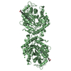
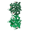
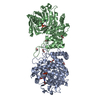
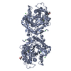
- PDB-3hm7: Crystal structure of allantoinase from Bacillus halodurans C-125 -
+
Open data
ID or keywords:
Loading...
-
Basic information
Entry
Database: PDB / ID: 3hm7
Title
Crystal structure of allantoinase from Bacillus halodurans C-125
Components
Allantoinase
Keywords
HYDROLASE / ALLANTOINASE / METALLO-DEPENDENT HYDROLASE / PROTEIN STRUCTURE INITIATIVE / PSI-2 / NEW YORK STRUCTURAL GENOMIX RESEARCH CONSORTIUM / NYSGXRC / STRUCTURAL GENOMICS / DNA-BINDING / Metal-binding / Purine metabolism / New York SGX Research Center for Structural Genomics
Function / homology
Function and homology information
allantoinase / allantoin catabolic process / allantoinase activity / purine nucleobase catabolic process / cobalt ion binding / zinc ion binding / cytoplasm Similarity search - Function
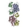
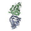
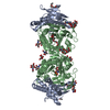
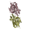
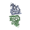
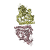
AUTHOR STATES THAT THE ASSEMBLY IS LIKELY A TETRAMER. THE ASU CONTAINS 1 FULL TETRAMER - MONOMERS A,B,E,F AND ANOTHER HALF (MONOMERS C AND D). THE REASON - AT LEAST TWO SIMILAR ENZYMES HAVE SIMILAR ASSEMBLIES DESPITE DIFFERENT CRYSTAL PACKING. IT WAS NOT EXPERIMENTALLY VERIFIED, HOWEVER.
-
Components
#1: Protein
Allantoinase / Allantoin-utilizing enzyme
Mass: 49818.484 Da / Num. of mol.: 6 Source method: isolated from a genetically manipulated source Source: (gene. exp.) Bacillus halodurans (bacteria) / Gene: allB, pucH, BH2309 / Production host: Escherichia coli (E. coli) / References: UniProt: Q9KAH8, allantoinase
In the structure databanks used in Yorodumi, some data are registered as the other names, "COVID-19 virus" and "2019-nCoV". Here are the details of the virus and the list of structure data.
Jan 31, 2019. EMDB accession codes are about to change! (news from PDBe EMDB page)
EMDB accession codes are about to change! (news from PDBe EMDB page)
The allocation of 4 digits for EMDB accession codes will soon come to an end. Whilst these codes will remain in use, new EMDB accession codes will include an additional digit and will expand incrementally as the available range of codes is exhausted. The current 4-digit format prefixed with “EMD-” (i.e. EMD-XXXX) will advance to a 5-digit format (i.e. EMD-XXXXX), and so on. It is currently estimated that the 4-digit codes will be depleted around Spring 2019, at which point the 5-digit format will come into force.
The EM Navigator/Yorodumi systems omit the EMD- prefix.
Related info.:Q: What is EMD? / ID/Accession-code notation in Yorodumi/EM Navigator
Yorodumi is a browser for structure data from EMDB, PDB, SASBDB, etc.
This page is also the successor to EM Navigator detail page, and also detail information page/front-end page for Omokage search.
The word "yorodu" (or yorozu) is an old Japanese word meaning "ten thousand". "mi" (miru) is to see.
Related info.:EMDB / PDB / SASBDB / Comparison of 3 databanks / Yorodumi Search / Aug 31, 2016. New EM Navigator & Yorodumi / Yorodumi Papers / Jmol/JSmol / Function and homology information / Changes in new EM Navigator and Yorodumi
 Movie
Movie Controller


 Open data
 Basic information
 Components
 Keywords
 Function and homology information
 Bacillus halodurans (bacteria)
 X-RAY DIFFRACTION /
  Authors
 Citation
 Structure visualization
 Downloads & links
 Other downloads
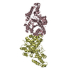

 PDBj
PDBj

 Assembly
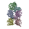

 Mass: 65.409 Da / Num. of mol.: 6 / Source method: obtained synthetically / Formula: Zn
 Mass: 18.015 Da / Num. of mol.: 371 / Source method: isolated from a natural source / Formula: H2O
 Sample preparation
 / Beamline: 31-ID / Wavelength: 0.9793
  Processing